. 2016 Oct;7(5):457.
doi: 10.4172/2155-9899.1000457. Epub 2016 Sep 26.

ER Stress-induced Inflammasome Activation Contributes to Hepatic Inflammation and Steatosis

Affiliations

ER Stress-induced Inflammasome Activation Contributes to Hepatic Inflammation and Steatosis

Jinyu Zhang et al. J Clin Cell Immunol. 2016 Oct.

Abstract

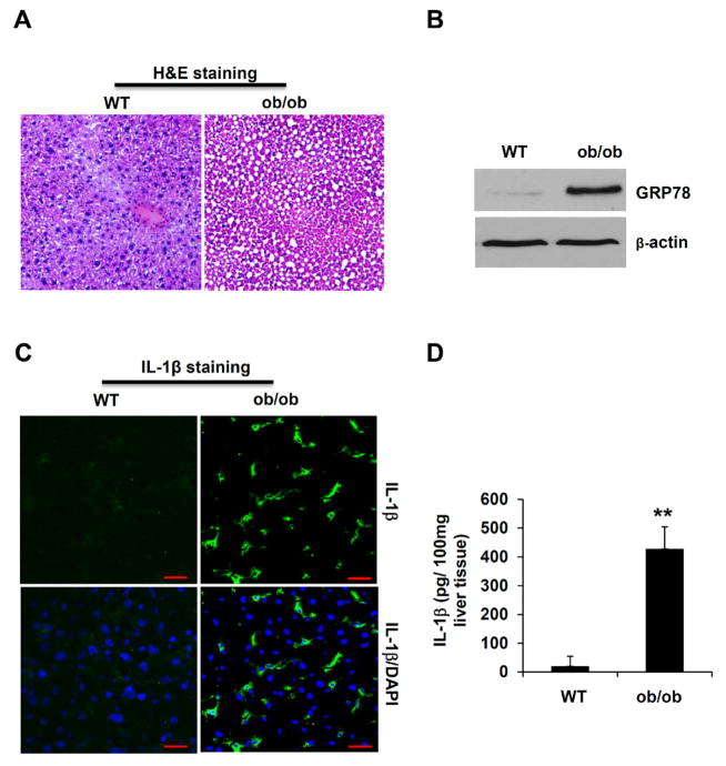
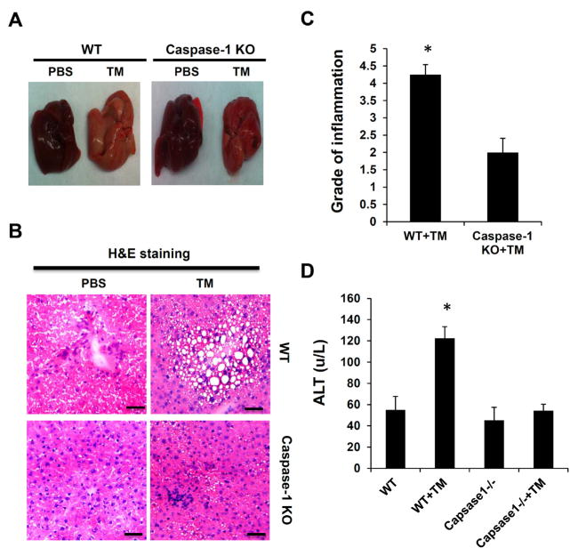
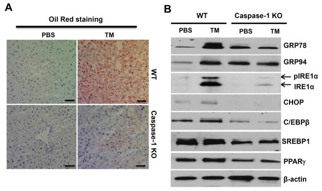
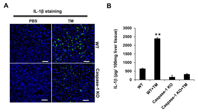
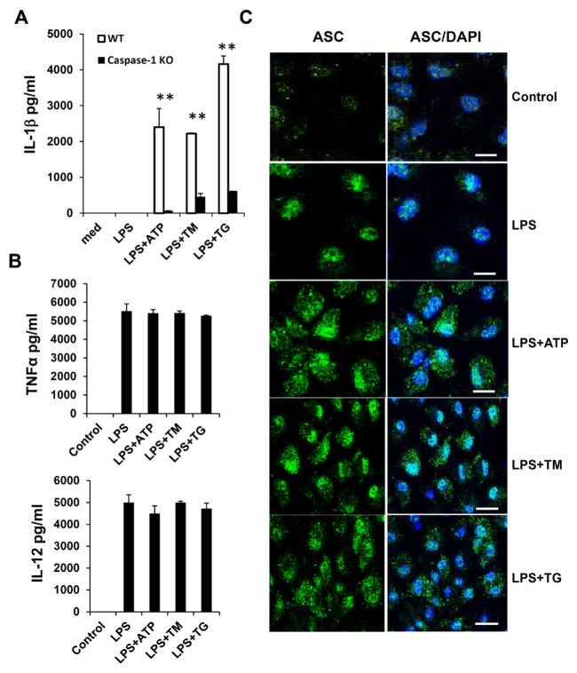
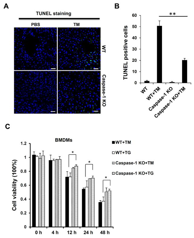
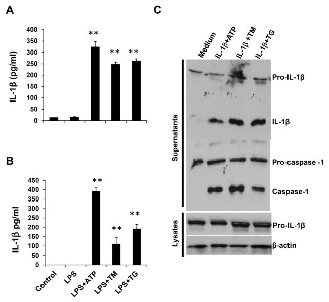
Endoplasmic reticulum (ER) stress functions as a protein folding and quality control mechanism to maintain cell homeostasis. Emerging evidence indicates that ER stress is also involved in metabolic and inflammatory diseases. However, the link between ER stress and inflammation remains not well characterized. In this study, we have demonstrated that ER stress-induced inflammasome activation plays a critical role in the pathogenesis of hepatic steatosis. By utilizing genetic and pharmacological agent-induced hepatic steatosis animal models, we found that hepatic steatosis was associated with inflammasome activation and ER stress. Our results show that caspase-1 ablation alleviated liver inflammation and injury. Liver tissues from caspase-1 KO mice had significantly reduced production of IL-1β under ER stress conditions. We also found that ER stress promoted inflammasome activation and IL-1β processing in both hepatocytes and Kupffer cells/macrophages. Moreover, lack of caspase-1 ameliorated cell death or pyropoptosis of hepatocytes induced by ER stress. Taken together, our findings suggest that ER stress-induced inflammasome activation and IL-1β production generate a positive feedback loop to amplify inflammatory response, eventually leading to liver steatosis and injury.

Keywords: Endoplasmic reticulum; Inflammasome; Liver; Steatosis.


Figures

Figure 1
Figure 1
The hepatic steatosis in the liver of ob/ob mice is associated with ER stress and inflammasome activation. (A) H&E staining of liver tissue sections from WT and ob/ob mice. (B) Liver tissues from WT and ob/ob mice were homogenized, and analyzed by Western Blot for ER stress markers Grp78. β-actin was used as a loading control. (C) Liver sections from WT and ob/ob mice were immunostained with an anti-IL-1β specific antibody. The green color indicates IL-1β, and nuclei are stained in blue by DAPI. Scale bar: 100 μm. (D) Liver tissues from WT and ob/ob mice were homogenized, and analyzed by IL-1β specific ELISA. Statistical significance is indicated, **P<0.01 (Student’s t test).
Figure 2
Figure 2
Caspase-1 deficiency attenuates ER stress-induced hepatic steatosis. Mice were intraperitoneally injected with TM (1 mg/kg body weight). (A) The morphology of liver, (B) H&E staining of liver tissue sections, (C) Pathologic inflammation scores, (D) plasma Alanine aminotransferase (ALT) level in liver tissues from WT and caspase-1 KO mice treated with or without TM. Each experiment was performed independently at least three times. *P<0.05, **P<0.01 (Student’s t test).
Figure 3
Figure 3
Inflammasomes regulate ER stress-induced lipogenesis. (A) Oil red O staining of liver tissue sections from WT and caspase-1 KO mice treated with or without TM. (B) Liver tissues from WT and caspase-1 KO mice treated with TM were homogenized, and analyzed by Western Blot for ER stress markers including Grp78, Grp94, CHOP and IRE1α, as well as lipogenic proteins such as C/EBPβ, PPARγ and SEBP1. β-actin was used as a loading control. Each experiment was performed independently at least three times.
Figure 4
Figure 4
ER stress-induced hepatic steatosis is associated with inflammasome activation and IL-1β production. (A) IL-1β immunostaining of liver sections. IL-1β levels in liver tissues from WT and caspase-1KO mice treated with or without TM were detected by immunostaining with an anti-IL-1β specific antibody. The green color indicates IL-1β, and nuclei are stained in blue by DAPI. Scale bar: 100 μm. (B) Liver tissues from WT and caspase-1 KO mice treated with TM treatment were homogenized, and analyzed by ELISA. Data are presented as means ± SEM. **P<0.01 (Student’s t test).
Figure 5
Figure 5
ER stress induces inflammasome activity and IL-1β processing in macrophages. (A) Bone marrow-derived macrophages (BMDMs) from WT and caspase-1 KO mice were primed with 250 ng/ml LPS for 4 hr and then treated with 1 mM ATP, 10 μg/ml TM or 5 μM TG. The concentrations of IL-1β in culture supernatants were assayed by ELISA. (B) The concentration of TNFα or IL-12 in culture supernatants of WT macrophages treated with LPS and ER stress inducers as in (A) was assayed by ELISA respectively. (C) Macrophages were treated as in (A), and then NLRP/ASC/caspase-1 complexes were detected using an anti-ASC specific antibody by immunostaining. The green color indicates the expression of ASC protein, and nuclei are stained in blue by DAPI. Scale bar: 100 μm. Data are presented as means ± SEM. **P<0.01 (Student’s t test).
Figure 6
Figure 6
Caspase-1 ablation alleviates cellular pyroptosis induced by ER stress. (A) TUNEL staining of liver sections from WT and caspase-1 KO mice injected with TM (n ≥ 5 for each group). The green color indicates TUNEL positive cell, and nuclei are stained in blue by DAPI. (B) The percentage of TUNEL positive cells in liver tissues from mice treated in (A). Liver from WT mice treated with TM displayed significantly more TUNEL positive cells throughout the tissue with several dense areas of TUNEL positive cells, indicating pyroptotic cell death. (C) Cell viabilities of TM- or TG-treated macrophages from WT and caspase-1 KO mice were examined by MTT (3-(4,5-dimethylthiazol-2-yl)-2,5-diphenyltetrazolium bromide) assay. Data are presented as means ± SEM. *P<0.05. **P<0.01 (Student’s t test).
Figure 7
Figure 7
ER stress induces inflammasome activation in both hepatocytes and Kupffer cells. Hepatocytes (A) and Kupffer cells (B) isolated from WT mice were primed with LPS, and then treated with ATP, TM or TG. IL-1β levels in culture supernatants were analyzed by ELISA. Data are presented as means ± SEM. **P<0.01 (Student’s t test). (C) IL-1β amplifies inflammasome activation and its own production. BMDMs from WT mice were pre-treated with IL-1, and then stimulated with ATP, TM or TG, the processing of IL-1β and caspase-1 in culture supernatants and lysates were analyzed by Western Blot.
Figure 8
Figure 8
ER stress induces inflammasome activation. ER stress triggers the Unfolded Protein Response (UPR), which is an adaptive response for stressed cells to retain ER homeostasis. However, dysregulated UPR can cause cell death and tissue damage. ER stress potentially induces the activation of inflammasomes and production of IL-1β, which in turn further exacerbates ER stress and inflammation. Inflammasome activation, together with UPR response, contributes to liver damage and steatosis.

References

    1. Kaufman RJ. Regulation of mRNA translation by protein folding in the endoplasmic reticulum. Trends Biochem Sci. 2004;29:152–158. - PubMed
    1. Castillo K, Rojas-Rivera D, Lisbona F, Caballero B, Nassif M, et al. BAX inhibitor-1 regulates autophagy by controlling the IRE1 branch of the unfolded protein response. EMBO J. 2011;30:4465–4478. - PMC - PubMed
    1. Schröder M, Kaufman RJ. The mammalian unfolded protein response. Annu Rev Biochem. 2005;74:739–789. - PubMed
    1. Wu J, Kaufman RJ. From acute ER stress to physiological roles of the Unfolded Protein Response. Cell Death Differ. 2006;13:374–384. - PubMed
    1. Xu C, Bailly-Maitre B, Reed JC. Endoplasmic reticulum stress: cell life and death decisions. J Clin Invest. 2005;115:2656–2664. - PMC - PubMed

LinkOut - more resources