

Altered structural and functional thalamocortical networks in secondarily generalized extratemporal lobe seizures
- PMID: 27942447
- PMCID: PMC5133650
- DOI: 10.1016/j.nicl.2016.11.010
Altered structural and functional thalamocortical networks in secondarily generalized extratemporal lobe seizures
Abstract
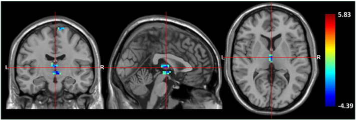
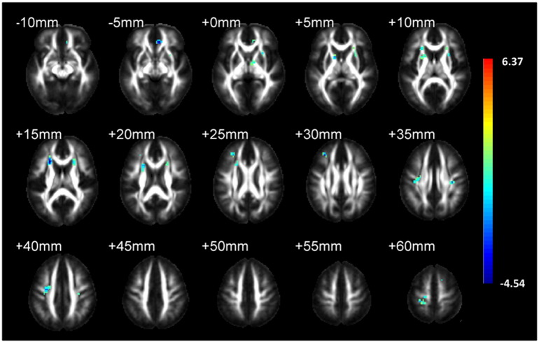
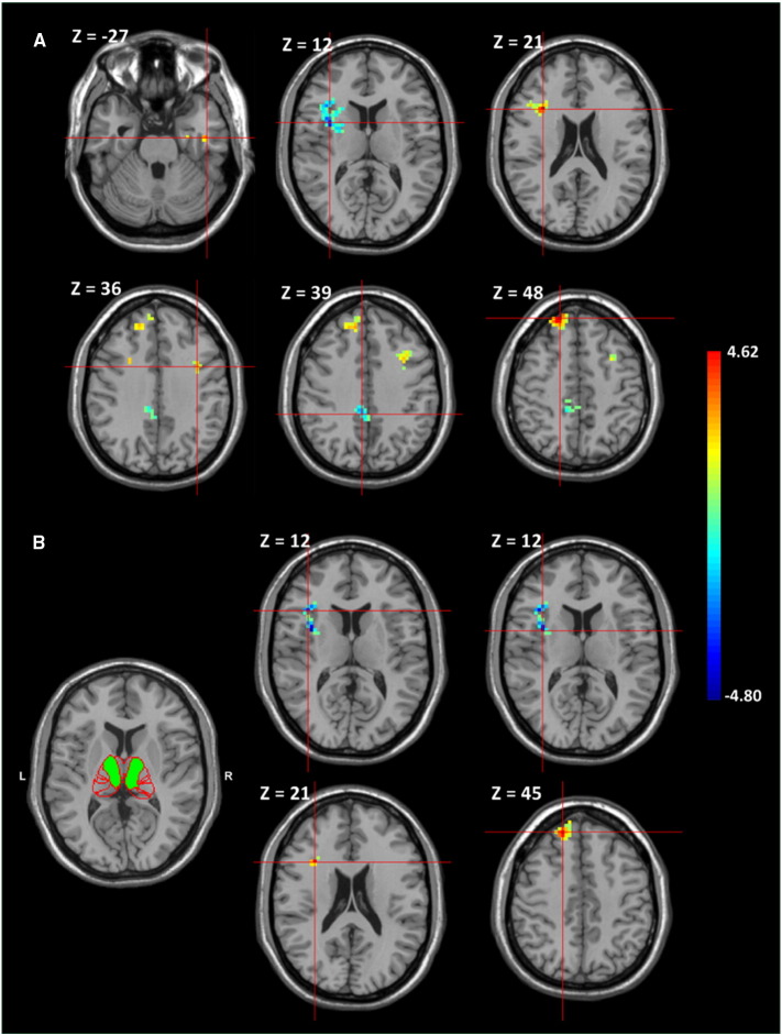
Structural and functional abnormalities in the thalamocortical network in primary generalized epilepsies or mesial temporal lobe epilepsy have recently been identified by voxel-wise analyses of neuroimaging. However, evidence is needed regarding the profiles of the thalamocortical network in patients with secondarily generalized seizures from focal neocortical sources. We used high-resolution T1-weighted, diffusion-tensor and resting-state functional MR imaging (rs-fMRI) to examine 16 patients with secondarily generalized extratemporal lobe seizures and 16 healthy controls. All the patients were medically effective and MRI-negative. Using whole brain voxel-based morphometry (VBM) to compare the patients with the normal controls, we observed significantly decreased gray matter (GM) density in the thalamus and 3 frontal gyri and significantly reduced white matter (WM) fractional anisotropy (FA) in the bilateral anterior corona radiata of the patients. Alterations in the thalamocortical functional connectivity with different cortices were identified by the rs-fMRI analysis seeding of the whole thalamus. The prefrontal gyri with the greatest functional connectivity were also traced by seeding a sub-thalamic region that is demarcated in an atlas, in which the thalamic parcellation is based on the WM connectivity to the cortices. This sub-thalamic region anatomically contains the mediodorsal thalamic nucleus where, concordantly, there was a significant decrease in thalamic GM density in the VBM study. In contrast to the negative correlation between the disease duration and reduced thalamic densities and subcortical FA values, the strength of the functional thalamocortical connectivity had a paradoxical correlation. Our results conclusively indicate that generalized seizures with a focal cortical source are associated with structural and functional alterations in the thalamocortical network.
Keywords: Fractional anisotropy; Generalized seizure; Gray matter density; Thalamocortical network.
Figures

References
-
- Alvim M.K., Coan A.C., Campos B.M., Yasuda C.L., Oliveira M.C., Morita M.E., Cendes F. Progression of gray matter atrophy in seizure-free patients with temporal lobe epilepsy. Epilepsia. 2016;57:621–629. - PubMed
-
- Arfanakis K., Hermann B.P., Rogers B.P., Carew J.D., Seidenberg M., Meyerand M.E. Diffusion tensor MRI in temporal lobe epilepsy. Magn. Reson. Imaging. 2002;20:511–519. - PubMed
-
- Ashburner J. A fast diffeomorphic image registration algorithm. NeuroImage. 2007;38:95–113. - PubMed
-
- Ashburner J., Friston K.J. Voxel-based morphometry–the methods. NeuroImage. 2000;11:805–821. - PubMed
-
- Ashburner J., Friston K.J. Unified segmentation. NeuroImage. 2005;26:839–851. - PubMed
Publication types
MeSH terms
LinkOut - more resources
Full Text Sources
Other Literature Sources
Medical
