

Posterior fossa syndrome-a narrative review
- PMID: 27942479
- PMCID: PMC5130561
- DOI: 10.21037/qims.2016.10.12
Posterior fossa syndrome-a narrative review
Retraction in
-
Retraction to posterior fossa syndrome-a narrative review.Quant Imaging Med Surg. 2017 Feb;7(1):169. doi: 10.21037/qims.2016.12.08. Quant Imaging Med Surg. 2017. PMID: 28275575 Free PMC article.
Abstract
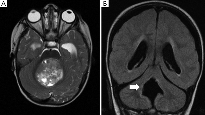
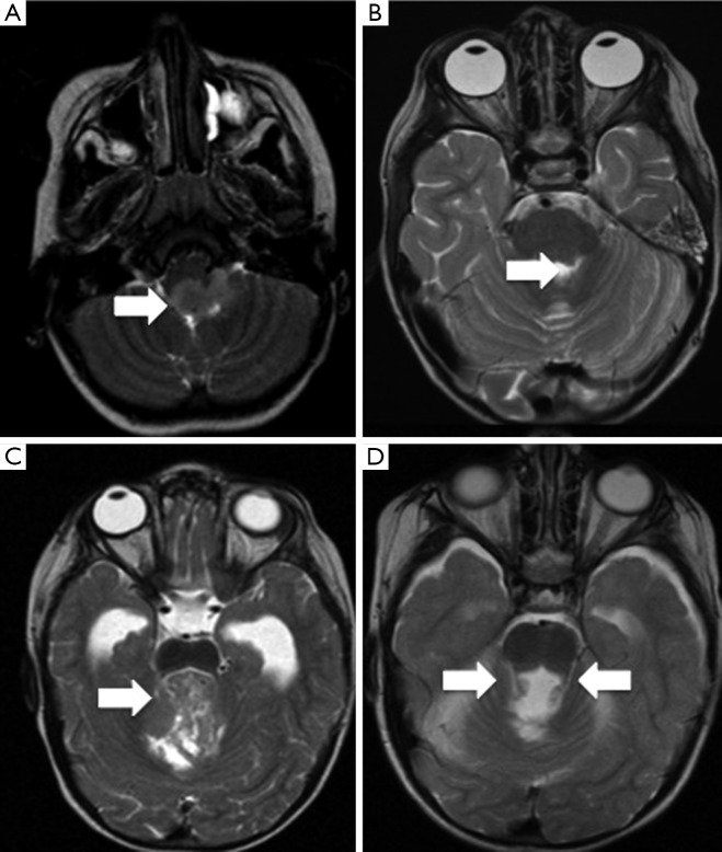
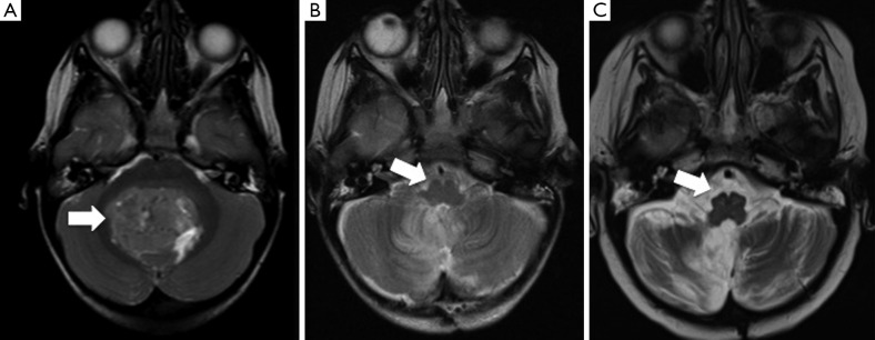
Posterior fossa syndrome (PFS), or cerebellar mutism syndrome (CMS), is a collection of neurological symptoms that occur following surgical resection of a posterior fossa tumour, and is characterised by either a reduction or an absence of speech. Some authors suggest that CM is only one symptom of the CMS complex that also includes ataxia, hypotonia and irritability as well as cranial nerve deficits, neurobehavioral changes and urinary retention or incontinence. It is seen almost exclusively in children. In 1985 Rekate et al. published the first work describing CM as a clinical entity, occurring as a consequence of bilateral cerebellar injury. Other associated symptoms include visual impairment, altered mood, impaired swallowing and significant gross and fine motor deficits. The effects of this can have a devastating impact on both the patient and their carers, posing a significant clinical challenge to neurorehabilitation services. The reported incidence was between 8% and 31% of children undergoing surgery for posterior fossa tumour. The underlying pathologies include vasospasm, oedema, and axonal/neuronal injury. Neuroimaging has contributed to a better understanding of the anatomical location of postoperative injury. There have been a number of suggestions for treatment interventions for PFS. However, apart from some individual reports, there have been no clinical trials indicating possible benefit. Occupational therapy, speech and language therapy, as well as neurocognitive support, contribute to the recovery of these patients.
Keywords: Posterior fossa syndrome (PFS); cerebellar mutism syndrome (CMS); neuroimaging; posterior fossa tumour.
Conflict of interest statement
The authors have no conflicts of interest to declare.
Figures
References
-
- De Smet HJ, Baillieux H, Catsman-Berrevoets C, De Deyn PP, Mariën P, Paquier PF. Postoperative motor speech production in children with the syndrome of 'cerebellar' mutism and subsequent dysarthria: a critical review of the literature. Eur J Paediatr Neurol 2007;11:193-207. 10.1016/j.ejpn.2007.01.007 - DOI - PubMed
Publication types
LinkOut - more resources
Full Text Sources
Other Literature Sources