. 2016 Dec 8;1(20):e89289.
doi: 10.1172/jci.insight.89289.

Tumor-infiltrating lymphocytes are dynamically desensitized to antigen but are maintained by homeostatic cytokine

Affiliations

Tumor-infiltrating lymphocytes are dynamically desensitized to antigen but are maintained by homeostatic cytokine

Bijan Boldajipour et al. JCI Insight. .

Abstract

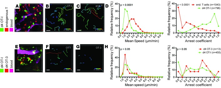
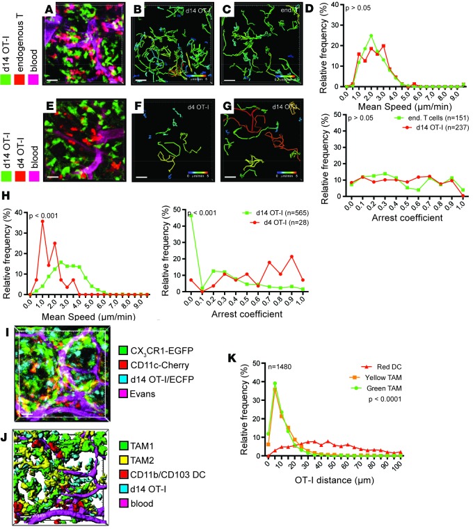
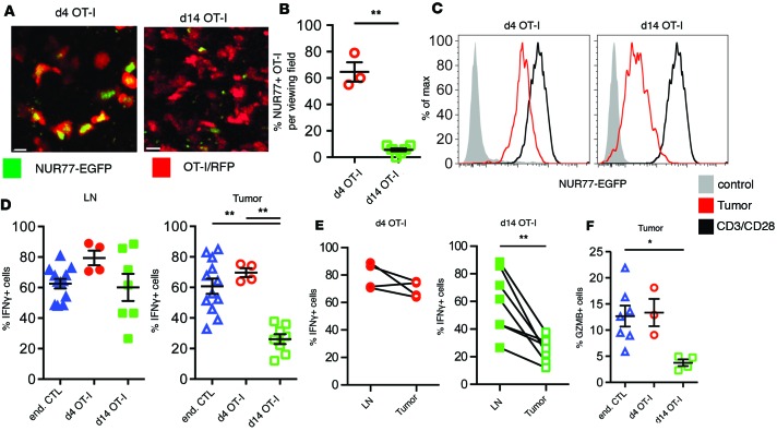
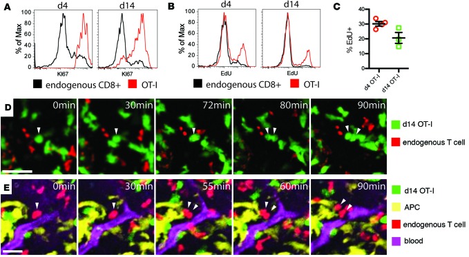
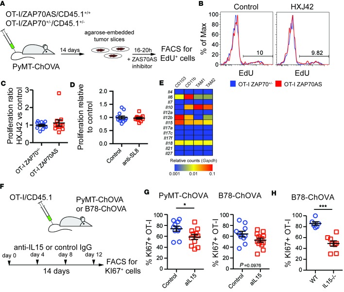
T cells that enter tumors are largely tolerized, but how that process is choreographed and how the ensuing "dysfunctional" tumor-infiltrating lymphocytes (TILs) are maintained are poorly understood and are difficult to assess in spontaneous disease. We exploited an autochthonous model of breast cancer for high-resolution imaging of the early and later stages of tumor residence to understand the relationships between cellular behaviors and cellular phenotypes. "Dysfunctional" differentiation began within the first days of tumor residence with an initial phase in which T cells arrest, largely on tumor-associated macrophages. Within 10 days, cellular motility increased and resembled a random walk, suggesting a relative absence of TCR signaling. We then studied the concurrent and apparently contradictory phenomenon that many of these cells express molecular markers of activation and were visualized undergoing active cell division. We found that whereas proliferation did not require ongoing TCR/ZAP70 signaling, instead this is driven in part by intratumoral IL-15 cytokine. Thus, TILs undergo sequential reprogramming by the tumor microenvironment and are actively retained, even while being antigen insensitive. We conclude that this program effectively fills the niche with ineffective yet cytokine-dependent TILs, and we propose that these might compete with new clones, when they arise.


Conflict of interest statement

The authors have declared that no conflict of interest exists.

Figures

Figure 1
Figure 1. T cell arrest upon arrival to the tumor is independent of TCR affinity.
(A–D) Migration parameters of high-affinity T cells in tumors upon arrival at the tumor site compared to endogenous T cells. (A–C) Representative video snapshot and migratory paths of high-affinity T cells and endogenous T cells over 2 hours, color-coded for mean track speed. CD11cYFP channel not displayed for ease of projection (see also Supplemental Video 1). (D) Kolmogorov-Smirnov analysis of the distribution of average migration speed and arrest coefficients. Data are representative of 6 experiments. (E–H) Migration parameters of high-affinity OT-I T cells compared with low-affinity OT-3 T cells upon arrival at the tumor site. (E–G) Representative video snapshot and migratory paths of high-affinity OT-I T cells and low-affinity OT-3 T cells over 2 hours, color-coded for mean track speed. (H) Kolmogorov-Smirnov analysis of the distribution of average migration speed and arrest coefficients. Due to the low numbers of OT-3 cells, data are pooled from 3 experiments. Scale bars: 20 μm.
Figure 2
Figure 2. Exposure to tumor environment alters T cell behavior.
(A–D) Migration parameters of tumor-resident high-affinity T cells compared with endogenous T cells. (A–C) Representative video snapshot and migratory paths of high-affinity T cells and endogenous T cells, color-coded for mean track speed over 2 hours. (D) Kolmogorov-Smirnov analysis of the distribution of average migration speed and arrest coefficients. Data are representative of 5 experiments. (E–H) Comparison of migration parameters of high-affinity T cells upon recent arrival to tumors (red) and after establishing residence (green) within the same animal. (EG) Representative video snapshot and migratory paths of high-affinity T cells upon recent arrival to tumors and after establishing residence, color-coded for mean track speed over 2 hours. (H) Kolmogorov-Smirnov analysis of the distribution of average migration speed and arrest coefficients. Data are representative of 5 experiments. (I) Snapshot and rendering (J) of the localization of high-affinity T cells (blue), CD11b+ TAM1 macrophages (green), CD11c+ TAM2 macrophages (yellow), and dendritic cells (red) 14 days after transfer of OT-I/CFP T cells into PyMT-ChOVA/CX3CR1-EGFP/CD11c-RFP transgenic hosts. Blood vessels were labeled by Evans Blue. (K) Representative statistical analysis of the distance of OT-I T cells to the next available APC using a Kruskal-Wallis 1-way ANOVA test. Data are representative of 2 experiments, n denotes the numbers of T cells analyzed. Scale bars: 20 μm.
Figure 3
Figure 3. Reduced TCR signaling and effector function in tumor-specific T cells.
(A) Representative images of OT-I/CD2-RFP/NUR77-EGFP reporter T cells after arrival and after establishing residence in tumors. Scale bars: 10 μm. (B) Statistical analysis of the percentage of NUR77-positive T cells in PyMT-ChOVA tumors using a Mann-Whitney t test, **P < 0.01. Data are analyzed from multiple tumors analyzed from 2 animals. (C) Analysis of the NUR77-EGFP reporter activity in OT-I T cells isolated from tumors (red). In vitro–activated T cells cultured for 24 hours on CD3/CD28-coated plates serve as positive control (black). OT-I/NUR77-EGFP T cells isolated from spleens of naive mice serve as negative control (filled gray histogram). Data are representative of at least 3 experiments. (D–F) Analysis of cytokine expression by ex vivo–restimulated CD44hi endogenous CD8+ T cells and OT-I T cells at different time points after adoptive transfer. (D) Cytokine expression in tumor-draining lymph nodes and tumors of PyMT-ChOVA animals. Statistical analysis was performed using a 1-way ANOVA Kruskal-Wallis test. (E) Comparison of cytokine expression in tumor-draining lymph nodes and their corresponding tumors. Samples were analyzed using the Wilcoxon matched-pairs rank test. (F) Granzyme B expression in tumor-resident T cells. Statistical analysis was performed using a 1-way ANOVA Kruskal-Wallis test, *P < 0.05, **P < 0.01.
Figure 4
Figure 4. Tumor-resident T cells proliferate in the tumor microenvironment.
(A and B) Expression of KI67 (A) and incorporation of EdU during DNA synthesis within 24 hours (B) in endogenous CD44hi CD8+ T cells (black), or high-affinity T cells (red), after arrival of OT-I T cells to the tumor (left) and after establishing residence (right). (C) Statistical analysis of EdU incorporation in several experiments. Mann-Whitney test, P > 0.05. (D and E) Snapshots of time-lapse videos of dividing high-affinity T cells (D, green) and endogenous T cells (E, red) in tumors of PyMT-ChOVA/CD2-RFP hosts. (E) CD11c+ antigen-presenting cells (APC, yellow) are visualized by the expression of an additional CD11c-YFP transgene, and blood vessels are labeled by Evans Blue (magenta). Arrows show dividing T cells and their daughter cells. Scale bar: 20 μm.
Figure 5
Figure 5. Proliferation of tumor-resident T cells is independent of TCR signaling but partially dependent on IL-15 signaling.
(A) Schematic of experiments to address TCR dependence of the proliferation of tumor-resident T cells. Genetically marked OT-I T cells expressing either a single copy of wild-type ZAP70 or the HXJ42-sensitive mutant ZAP70AS were transferred to PyMT-ChOVA mice. Fourteen days later, tumor slice cultures were prepared from tumors and incubated for 24 hours in the presence of EdU and either HXJ42 inhibitor or DMSO control. (B) Representative analysis of EdU incorporation of inhibitor-resistant OT-I/ZAP70+/– or inhibitor-sensitive OT-I/ZAP70AS T cells in the presence or absence of the inhibitor using flow cytometry. (C) The ratio of proliferation in tumor slices incubated with HXJ42 inhibitor over the proliferation in control slices was analyzed using a Mann-Whitney test. P > 0.05. Data are pooled from 3 experiments. (D) Similar to A–C, tumor slices of PyMT-ChOVA mice treated with OT-I T cells were incubated with EdU in absence or presence of an antibody that blocks the MHCI-presented ovalbumin peptide (anti-SL8). EdU incorporation was compared against the average of the control group using a Mann-Whitney test. P > 0.05. Data are pooled from 3 experiments. (E) Analysis of homeostatic cytokine expression by myeloid cells in the tumor microenvironment. Cytokine RNA expression data from ref. were mined for each indicated cell type and plotted using the indicated color scale and using Gapdh expression for normalization. (F) Schematic of experiments addressing the influence of IL-15 signaling on T cell proliferation in two tumor models. KI67+ OT-I cells served as a readout of cell proliferation. (G) Statistical analysis of KI67 expression of tumor-resident OT-I T cells in B78-ChOVA and PyMT-ChOVA tumors treated with IL-15-blocking antibody or control using a Mann-Whitney test. *P < 0.05. Data are pooled from 2 experiments. (H) Statistical analysis of KI67 expression of tumor-resident OT-I T cells in B78-ChOVA tumors grown in wild-type or IL-15-deficient animals using a Mann-Whitney test. ***P < 0.001. Data are pooled from 2 experiments.

References

    1. Hellström I, Hellström KE, Pierce GE, Yang JP. Cellular and humoral immunity to different types of human neoplasms. Nature. 1968;220(5174):1352–1354. doi: 10.1038/2201352a0. - DOI - PubMed
    1. Wherry EJ, et al. Molecular signature of CD8+ T cell exhaustion during chronic viral infection. Immunity. 2007;27(4):670–684. doi: 10.1016/j.immuni.2007.09.006. - DOI - PubMed
    1. Baitsch L, et al. Exhaustion of tumor-specific CD8+ T cells in metastases from melanoma patients. J Clin Invest. 2011;121(6):2350–2360. doi: 10.1172/JCI46102. - DOI - PMC - PubMed
    1. Engelhardt JJ, et al. Marginating dendritic cells of the tumor microenvironment cross-present tumor antigens and stably engage tumor-specific T cells. Cancer Cell. 2012;21(3):402–417. doi: 10.1016/j.ccr.2012.01.008. - DOI - PMC - PubMed
    1. Gubin MM, et al. Checkpoint blockade cancer immunotherapy targets tumour-specific mutant antigens. Nature. 2014;515(7528):577–581. doi: 10.1038/nature13988. - DOI - PMC - PubMed

Publication types