Review
. 2017 May;74(9):1605-1624.
doi: 10.1007/s00018-016-2418-4. Epub 2016 Dec 10.

Complement factor H in host defense and immune evasion

Affiliations
Review

Complement factor H in host defense and immune evasion

Raffaella Parente et al. Cell Mol Life Sci. 2017 May.

Abstract

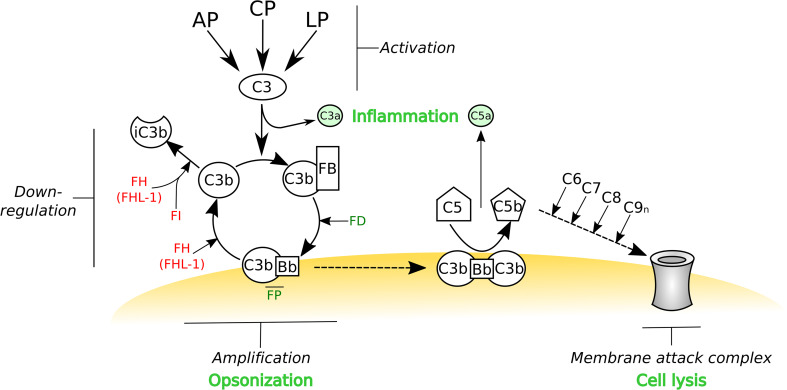
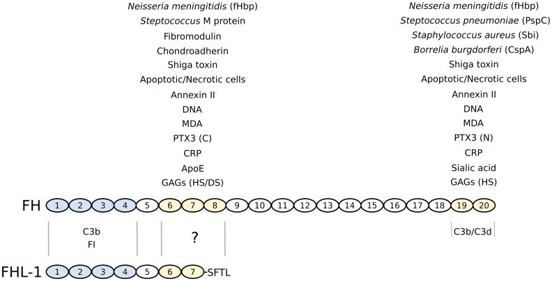
Complement is the major humoral component of the innate immune system. It recognizes pathogen- and damage-associated molecular patterns, and initiates the immune response in coordination with innate and adaptive immunity. When activated, the complement system unleashes powerful cytotoxic and inflammatory mechanisms, and thus its tight control is crucial to prevent damage to host tissues and allow restoration of immune homeostasis. Factor H is the major soluble inhibitor of complement, where its binding to self markers (i.e., particular glycan structures) prevents complement activation and amplification on host surfaces. Not surprisingly, mutations and polymorphisms that affect recognition of self by factor H are associated with diseases of complement dysregulation, such as age-related macular degeneration and atypical haemolytic uremic syndrome. In addition, pathogens (i.e., non-self) and cancer cells (i.e., altered-self) can hijack factor H to evade the immune response. Here we review recent (and not so recent) literature on the structure and function of factor H, including the emerging roles of this protein in the pathophysiology of infectious diseases and cancer.

Keywords: Cancer immunology; Complement cascade; Complement factor H; Glycan markers; Inflammatory diseases; Innate immunity.


Figures

Fig. 1
Fig. 1
The activation, amplification and regulation of the complement system. The complement system can be activated by three different pathways: the alternative pathway (AP), classical pathway (CP) and lectin pathway (LP). While the AP is constitutively active and undergoes a constant tick over, the CP and LP are triggered by antibody- and carbohydrate-mediated recognition mechanisms, respectively. Regardless of how this protein cascade is initiated, it is the AP that amplifies the complement system; e.g. if the AP C3 convertase is formed on an activator surface. All three pathways lead to the conversion of C3 into C3b, which can become covalently attached (via a thioester) to any nearby nucleophile (e.g., a hydroxyl or amine group on a surface); C3 cleavage releases the small protein fragment C3a, a potent anaphylatoxin. The remaining C3b can associate with factor B (FB), which is itself cleaved by factor D (FD). This forms the AP C3 convertase (i.e., C3bBb) where this is stabilized by factor P (FP). If this convertase is not deactivated then any free C3 in the vicinity will be converted into C3b and thus begins a positive feedback cycle, referred to as the amplification loop. If left to run unchecked, this will lead to the opsonization of the target surface (with C3b) and the formation of the AP C5 convertase (i.e., C3bBbC3b), which represents the initiation of the terminal pathway of complement and the subsequent formation of the membrane-attack complex (MAC) that can lead to lysis (e.g., of bacteria). Host cells have a number of cell surface proteins capable of down-regulating the complement cascade that can either dissociate the AP C3 convertase, known as decay acceleration activity, or act as cofactors for the proteolytic cleavage, by factor I (FI), to inactive C3b (iC3b). The soluble C3b-binding protein factor H (FH) can associate with host cells via the recognition of glycan markers of self and thereby down-regulate complement through its decay accelerating and cofactor activities. FH (and the truncated FHL-1 product of the CFH gene) can also recognize self markers on acellular structures such as the extracellular matrix, and in this context FH/FHL-1 are the only negative regulators of the complement AP pathway. Therefore, FH and FHL-1 have a pivotal role in the prevention of complement activation in host tissues. This schematic is modified from [184]
Fig. 2
Fig. 2
Domain structure and ligand-binding activities of factor H and FHL-1. Complement FH is comprised of twenty CCP modules, whereas FHL-1 shares the first seven of these followed by a unique four amino acid sequence (SFTL). The four N-terminal CCPs of FH/FHL-1 (colored blue) confer regulatory activity through mediating C3b and FI binding; C3b (via its C3d region) also binds to the C-terminal CCPs 19–20 of FH. The CCPs 6–8 and/or 19–20 regions (colored yellow) support the interactions with a wide variety of ligands including glycans (GAGs and sialic acid), pentraxins (CRP and PTX3; where the latter binds through the its C- and N-terminal domains, respectively), the lipid peroxidation product MDA, apoptotic/necrotic cells (e.g., via annexin II and DNA), extracellular matrix proteins (e.g., chondroadherin and fibromodulin), ApoE, shiga toxin from enterohaemorrhagic E. coli, and FH-binding proteins from various other bacteria. Although it is presumed that many of the ligands that bind FH via CCPs 6–8 will interact also with FHL-1, most of these have yet to be tested
Fig. 3
Fig. 3
A dual role for FH in cancer. FH is expressed or recruited by cancer cells and this leads to reduced deposition of C3 and less production of the anaphylatoxin C5a. Inhibition of complement attack (complement resistance) can favor tumor survival and promote tumor growth (tumor promotion). However, in certain types of cancer that are inherently promoted and sustained by inflammation, the anti-inflammatory microenvironment generated by FH (complement assistance) might be unfavorable to tumor growth and progression (tumor suppression)

References

    1. Bottazzi B, Doni A, Garlanda C, Mantovani A. An integrated view of humoral innate immunity: pentraxins as a paradigm. Annu Rev Immunol. 2010;28:157–183. doi: 10.1146/annurev-immunol-030409-101305. - DOI - PubMed
    1. Iwasaki A, Medzhitov R. Regulation of adaptive immunity by the innate immune system. Science. 2010;327(5963):291–295. doi: 10.1126/science.1183021. - DOI - PMC - PubMed
    1. Ricklin D, Reis ES, Lambris JD. Complement in disease: a defence system turning offensive. Nat Rev Nephrol. 2016;12(7):383–401. doi: 10.1038/nrneph.2016.70. - DOI - PMC - PubMed
    1. Kemper C, Pangburn MK, Fishelson Z. Complement nomenclature 2014. Mol Immunol. 2014;61(2):56–58. doi: 10.1016/j.molimm.2014.07.004. - DOI - PubMed
    1. Medzhitov R. Origin and physiological roles of inflammation. Nature. 2008;454(7203):428–435. doi: 10.1038/nature07201. - DOI - PubMed

Publication types