

Flow Cytometric Analysis of Extracellular Vesicles
- PMID: 27943218
- PMCID: PMC7888554
- DOI: 10.1007/978-1-4939-6728-5_16
Flow Cytometric Analysis of Extracellular Vesicles
Abstract
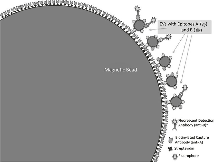
To analyze EVs with conventional flow cytometers, most researchers will find it necessary to bind EVs to beads that are large enough to be individually resolved on the flow cytometer available in their lab or facility. Although high-resolution flow cytometers are available and are being used for EV analysis, the use of these instruments for studying EVs requires careful use and validation by experienced small-particle flow cytometrists, beyond the scope of this chapter. Shown here is a method for using streptavidin-coated beads to capture biotinylated antibodies, and stain the bead-bound EVs with directly conjugated antibodies. We find that this method is a useful tool not only on its own, without further high resolution flow cytometric analysis, but also as a means for optimizing staining methods and testing new labels for later use in high resolution, single EV flow cytometric studies. The end of the chapter includes sphere-packing calculations to quantify aspects of EV- and bead-surface geometry, as a reference for use as readers of this chapter optimize their own flow cytometry assays with EVs.
Keywords: Exosomes; Extracellular vesicles; Flow cytometry; Subsets.
Figures


Similar articles
-
Flow cytometric analysis of extracellular vesicle subsets in plasma: impact of swarm by particles of non-interest.J Thromb Haemost. 2018 Jul;16(7):1423-1436. doi: 10.1111/jth.14154. Epub 2018 Jun 15. J Thromb Haemost. 2018. PMID: 29781099
-
Comparison of EV characterization by commercial high-sensitivity flow cytometers and a custom single-molecule flow cytometer.J Extracell Vesicles. 2024 Aug;13(8):e12498. doi: 10.1002/jev2.12498. J Extracell Vesicles. 2024. PMID: 39140467 Free PMC article.
-
Bead-Based Extracellular Vesicle Analysis Using Flow Cytometry.Adv Biosyst. 2020 Dec;4(12):e2000203. doi: 10.1002/adbi.202000203. Epub 2020 Oct 25. Adv Biosyst. 2020. PMID: 33103361 Free PMC article.
-
Detection of platelet vesicles by flow cytometry.Platelets. 2017 May;28(3):256-262. doi: 10.1080/09537104.2017.1280602. Epub 2017 Mar 2. Platelets. 2017. PMID: 28277059 Free PMC article. Review.
-
Methods to Analyze EVs.Methods Mol Biol. 2017;1545:1-20. doi: 10.1007/978-1-4939-6728-5_1. Methods Mol Biol. 2017. PMID: 27943203 Review.
Cited by
-
Exosomes in disease and regeneration: biological functions, diagnostics, and beneficial effects.Am J Physiol Heart Circ Physiol. 2020 Dec 1;319(6):H1162-H1180. doi: 10.1152/ajpheart.00075.2020. Epub 2020 Sep 28. Am J Physiol Heart Circ Physiol. 2020. PMID: 32986962 Free PMC article.
-
Proteomes of exosomes from HPV(+) or HPV(-) head and neck cancer cells: differential enrichment in immunoregulatory proteins.Oncoimmunology. 2019 Apr 15;8(7):1593808. doi: 10.1080/2162402X.2019.1593808. eCollection 2019. Oncoimmunology. 2019. PMID: 31143515 Free PMC article.
-
HIF-1α and Pro-Inflammatory Signaling Improves the Immunomodulatory Activity of MSC-Derived Extracellular Vesicles.Int J Mol Sci. 2021 Mar 26;22(7):3416. doi: 10.3390/ijms22073416. Int J Mol Sci. 2021. PMID: 33810359 Free PMC article.
-
Extracellular Vesicles for Research on Psychiatric Disorders.Schizophr Bull. 2019 Jan 1;45(1):7-16. doi: 10.1093/schbul/sby127. Schizophr Bull. 2019. PMID: 30239909 Free PMC article. Review.
-
Exosomes: Methods for Isolation and Characterization in Biological Samples.Methods Mol Biol. 2024;2835:181-213. doi: 10.1007/978-1-0716-3995-5_17. Methods Mol Biol. 2024. PMID: 39105917 Review.
References
-
- Erdbrugger U, Lannigan J (2016) Analytical challenges of extracellular vesicle detection: a comparison of different techniques. Cytometry A 89(2):123–134 - PubMed
MeSH terms
Substances
Grants and funding
LinkOut - more resources
Full Text Sources
Other Literature Sources
Miscellaneous
