

Histologic validation of myocardial fibrosis measured by T1 mapping: a systematic review and meta-analysis
- PMID: 27955698
- PMCID: PMC5154013
- DOI: 10.1186/s12968-016-0313-7
Histologic validation of myocardial fibrosis measured by T1 mapping: a systematic review and meta-analysis
Abstract
Background: Myocardial fibrosis is being increasingly recognised as a common final pathway of a wide range of diseases. Thus, the development of an accurate and convenient method to evaluate myocardial fibrosis is of major importance. Although T1 mapping is a potential alternative for myocardial biopsy, validation studies are limited to small numbers and vary regarding technical facets, and include only a restricted number of disease. A systematic review and meta-analysis was conducted to objectively and comprehensively evaluate the performance of T1 mapping on the quantification of myocardial fibrosis using cardiovascular magnetic resonance (CMR).
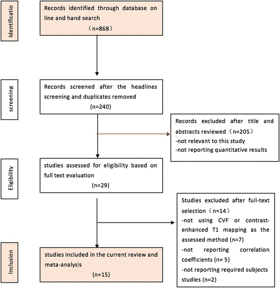
Methods: PubMed, EMBASE and the Cochrane Library databases were searched for studies applying T1 mapping to measure myocardial fibrosis and that validated the results via histological analysis. A pooled correlation coefficient between the CMR and histology measurements was used to evaluate the performance of the T1 mapping.
Results: A total of 15 studies, including 308 patients who had CMR and myocardial biopsy were included and the pooled correlation coefficient between ECV measured by T1 mapping and biopsy for the selected studies was 0.884 (95% CI: 0.854, 0.914) and was not notably heterogeneous chi-squared = 7.44; P = 0.489 for the Q test and I^2 = 0.00%).
Conclusions: The quantitative measurement of myocardial fibrosis via T1 mapping is associated with a favourable overall correlation with the myocardial biopsy measurements. Further studies are required to determine the calibration of the T1 mapping results for the biopsy findings of different cardiomyopathies.
Keywords: Cardiovascular magnetic resonance, T1 mapping; Myocardial fibrosis.
Figures


References
-
- Karamitsos TD, Francis JM, Myerson S, Selvanayagam JB, Neubauer S. The Role of Cardiovascular Magnetic Resonance Imaging in Heart Failure. Jac. 2009;54:1407–1424. - PubMed
-
- Bohl S, Wassmuth R, Abdel-Aty H, Rudolph A, Messroghli D, Dietz R, Schulz-Menger J. Delayed enhancement cardiac magnetic resonance imaging reveals typical patterns of myocardial injury in patients with various forms of non-ischemic heart disease. Int J Cardiovasc Imaging. 2008;24:597–607. doi: 10.1007/s10554-008-9300-x. - DOI - PubMed
-
- Dweck MR, Joshi S, Murigu T, Alpendurada F, Jabbour A, Melina G, Banya W, Gulati A, Roussin I, Raza S, Prasad NA, Wage R, Quarto C, Angeloni E, Refice S, Sheppard M, Cook SA, Kilner PJ, Pennell DJ, Newby DE, Mohiaddin RH, Pepper J, Prasad SK. Midwall fibrosis is an independent predictor of mortality in patients with aortic stenosis. J Am Coll Cardiol [Internet] 2011;58:1271–1279. doi: 10.1016/j.jacc.2011.03.064. - DOI - PubMed
-
- O’Hanlon R, Grasso A, Roughton M, Moon JC, Clark S, Wage R, Webb J, Kulkarni M, Dawson D, Sulaibeekh L, Chandrasekaran B, Bucciarelli-Ducci C, Pasquale F, Cowie MR, McKenna WJ, Sheppard MN, Elliott PM, Pennell DJ, Prasad SK. Prognostic significance of myocardial fibrosis in hypertrophic cardiomyopathy. J Am Coll Cardiol [Internet] 2010;56:867–874. doi: 10.1016/j.jacc.2010.05.010. - DOI - PubMed
Publication types
MeSH terms
LinkOut - more resources
Full Text Sources
Other Literature Sources
Medical
