. 2017 Jan;187(1):187-199.
doi: 10.1016/j.ajpath.2016.09.002. Epub 2016 Dec 9.

Salmonella Meningitis Associated with Monocyte Infiltration in Mice

Affiliations

Salmonella Meningitis Associated with Monocyte Infiltration in Mice

Timothy J Bauler et al. Am J Pathol. 2017 Jan.

Abstract

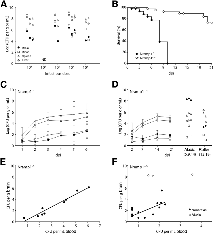
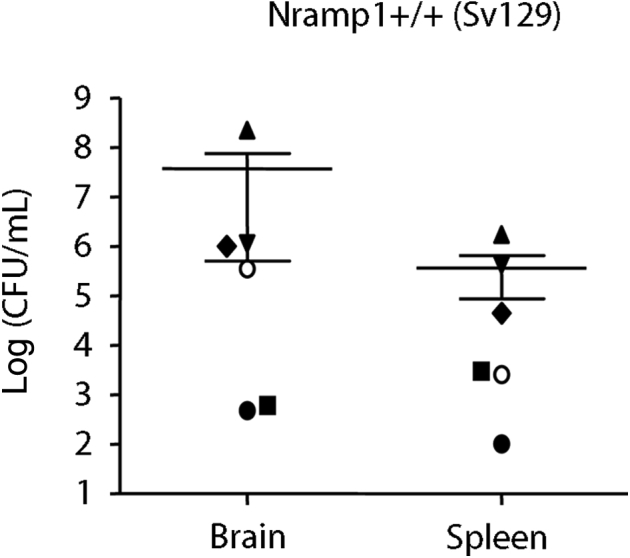
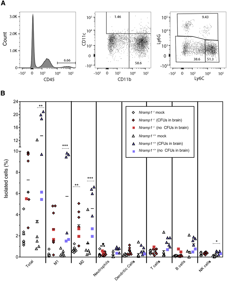
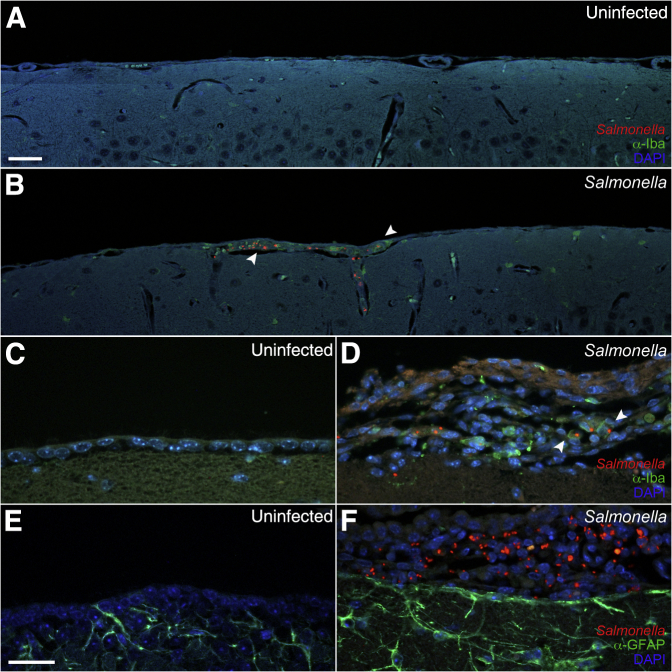
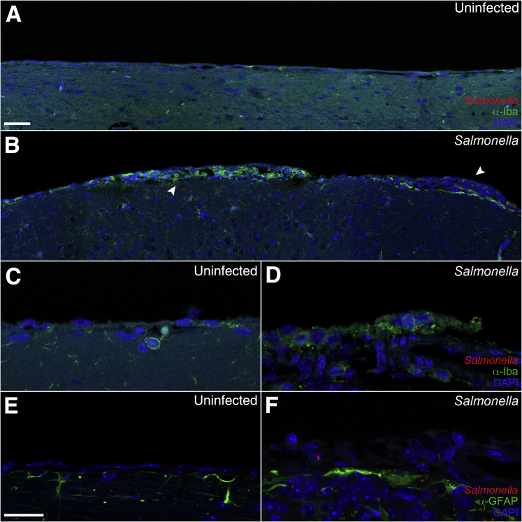
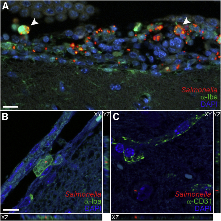
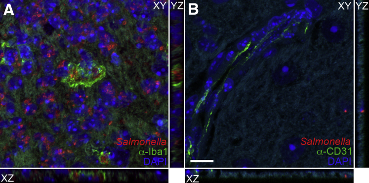
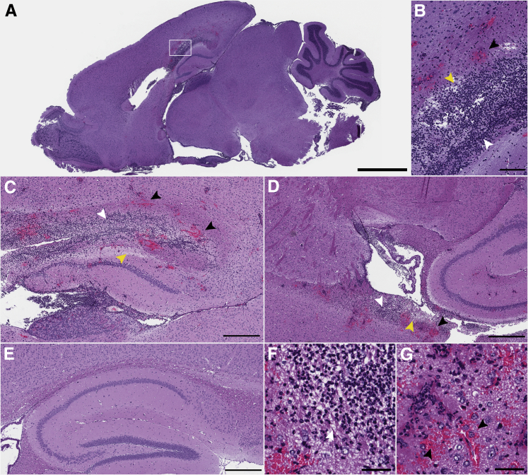
In the current study, we examined the ability of Salmonella enterica serovar Typhimurium to infect the central nervous system and cause meningitis following the natural route of infection in mice. C57BL/6J mice are extremely susceptible to systemic infection by Salmonella Typhimurium because of loss-of-function mutations in Nramp1 (SLC11A1), a phagosomal membrane protein that controls iron export from vacuoles and inhibits Salmonella growth in macrophages. Therefore, we assessed the ability of Salmonella to disseminate to the central nervous system (CNS) after oral infection in C57BL/6J mice expressing either wild-type (resistant) or mutant (susceptible) alleles of Nramp1. In both strains, oral infection resulted in focal meningitis and ventriculitis with recruitment of inflammatory monocytes to the CNS. In susceptible Nramp1-/- mice, there was a direct correlation between bacteremia and the number of bacteria in the brain, which was not observed in resistant Nramp1+/+ mice. A small percentage of Nramp1+/+ mice developed severe ataxia, which was associated with high bacterial loads in the CNS as well as clear histopathology of necrotizing vasculitis and hemorrhage in the brain. Thus, Nramp1 is not essential for Salmonella entry into the CNS or neuroinflammation, but may influence the mechanisms of CNS entry as well as the severity of meningitis.


Figures

Figure 1
Figure 1
Salmonella infection and dissemination in Nramp1−/− and Nramp1+/+ mice. A: Organ colonization of Nramp1−/− mice with clinical salmonellosis. Mice were infected orally with 104, 106, 107, or 108Salmonella. Samples were collected on development of clinical disease, up to 10 days postinfection (dpi). B: Percentage survival of Nramp1−/− and Nramp1+/+ mice infected with 108 or 109 colony-forming units (CFUs) Salmonella, respectively. C: Bacterial loads in Nramp1−/− mice infected with 108Salmonella. D: Bacterial loads in Nramp1+/+ mice infected with 109Salmonella. Mice with clinical ataxia or rolling disease are shown separately. E and F: Relationship between bacterial loads in the blood and brain of Nramp1−/− (E) or Nramp1+/+ (F) mice at 6 or 21 days postinfection. Data were fitted using a linear regression model of transformed log-log CFU data. Mice with ataxia are indicated. Data represent means ± SD (C and D). n = 3 (A, 104 and 107Salmonella); n = 2 (A, 106 and 108Salmonella); n = 50 (B, Nramp1−/−); n = 54 (B, Nramp1+/+); n = 3 to 14 mice per time point (C); n = 2 to 20 mice per time point (D).
Figure 2
Figure 2
Salmonella colonization of the brain of 129S6 (129SvEv) mice. Mice were orally infected with 109Salmonella. Samples were collected 2 weeks after infection. Each symbol represents one mouse. Means and SEM are shown. n = 6. CFU, colony-forming unit.
Figure 3
Figure 3
Infiltration of immune cells in the brains of infected Nramp1−/− and Nramp1+/+ mice. Mice were orally infected with Salmonella, and samples were collected on day 6 (Nramp1−/−) and day 14 (Nramp1+/+) after infection. A: Example of gated analysis used for determining M1 and M2 cell phenotypes. B: Percentage of infiltrating immune cells in the brains of infected Nramp1−/− (red symbols) and Nramp1+/+ (blue symbols) mice. Half of the brain was used to estimate the bacterial load [colony-forming units (CFUs)], whereas the other half was processed for flow cytometry. Infected mice, where no CFUs were detected, are indicated for Nramp1−/− (red squares) and Nramp1+/+ (blue squares). Mock-infected Nramp1−/− (white diamonds) and Nramp1+/+ (white triangles) mice were used as controls. Each symbol represents one mouse. n = 8 (B, infected and mock-infected Nramp1−/− mice); n = 6 (B, infected and mock-infected Nramp1+/+ mice). P < 0.05, ∗∗P < 0.01, and ∗∗∗P < 0.001. NK, natural killer.
Figure 4
Figure 4
Meningitis in Nramp1−/− mice after Salmonella infection. Brain tissue of Nramp1−/− mice orally infected with Salmonella was fixed and immunostained up to 10 days after infection. A and B: Extended representative images of uninfected (A) and infected (B) brain tissue immunostained for Iba1 (green), Salmonella (red), and DAPI (blue). Arrowheads indicate regions of meningitis. C–F: High-magnification representative images of uninfected (C and E) and infected (D and F) brain tissue immunostained for Salmonella (red), DAPI (blue), and either glial fibrillary acidic protein (GFAP) or Iba1 (green). Scale bars = 40 μm (AF).
Figure 5
Figure 5
Meningitis in Nramp1+/+ mice after Salmonella infection. Brain tissue of Nramp1+/+ mice orally infected with Salmonella was fixed and immunostained up to 21 days after infection. A and B: Extended representative images of uninfected (A) and infected (B) brain tissue immunostained for Iba1 (green), Salmonella (red), and DAPI (blue). Arrowheads indicate regions of meningitis. C–F: High-magnification representative images of uninfected (C and E) and infected (D and F) brain tissue immunostained for Salmonella (red), DAPI (blue), and either glial fibrillary acidic protein (GFAP) or Iba1 (green). Scale bars: 40 μm (A and B); 20 μm (CF).
Figure 6
Figure 6
Salmonella intracellular infection of the ventricle, meninges, and parenchyma. Brain tissue of Nramp1−/− mice orally infected with Salmonella was fixed and immunostained up to 10 days after infection. A: Representative image of infected ventricular brain tissue immunostained for Iba1 (green), Salmonella (red), and DAPI (blue). Arrowheads indicate examples of intracellular bacteria. B and C: XYZ projections illustrating presence of intracellular and extravascular bacteria in the meninges (B) and parenchyma (C) immunostained for Salmonella (red), DAPI (blue), and either Iba1 (green; B) or CD31 (green; C). XZ or YZ images were acquired in the XY plane with slices taken along the Z-axis. Scale bars: 20 μm (A); 10 μm (B and C).
Figure 7
Figure 7
Salmonella infection of the brain parenchyma in Nramp1+/+ neurological mice. Brain tissue of Nramp1+/+ mice orally infected with Salmonella was fixed and immunostained at 5, 9, and 14 days after infection. XYZ projections illustrating presence of intracellular and extravascular bacteria in the parenchyma immunostained for Salmonella (red), DAPI (blue), and either Iba1 (green; A) or CD31 (green; B). XZ and YZ images were acquired in the XY plane with slices taken along the Z-axis. Scale bar = 10 μm (A and B).
Figure 8
Figure 8
Neutrophil infiltration, focal hemorrhaging, and ventriculitis in the brains of ataxic Nramp1+/+ mice. Brains from mice with clinical signs of ataxia after infection with Salmonella were stained with hematoxylin and eosin. A: Low-magnification image showing neutrophil infiltration and hemorrhaging (boxed area), magnified in B. Malacia with neutrophil infiltrate and focal hemorrhaging in the corpus callosum (B and C) and lateral ventricle (D). E: Image of hippocampus and corpus callosum from an uninfected mouse for comparison. F and G: Infiltration and vascular thrombosis. Black arrowheads indicate regions of hemorrhage; yellow arrowheads, regions of tissue clearing; and white arrowheads, regions of cellular infiltrate. Scale bars: 2 mm (A); 200 μm (B); 500 μm (C–E); 50 μm (F and G).
Figure 9
Figure 9
Bacterial accumulation and macrophage infiltration in the brains of ataxic Nramp1+/+ mice. Brains from mice with clinical signs of ataxia after infection with Salmonella were immunostained. A: Low-magnification image of infected brain tissue immunostained for glial fibrillary acidic protein (GFAP; green), Salmonella (red), and DAPI (blue). Boxed regions illustrate bacteria in the corpus collosum, olfactory bulb, and meningies and are magnified in B, C, and F, respectively. For orientation purposes, the olfactory bulb (OB) and cerebellum (CB) are labeled. Representative image of Salmonella in the corpus callosum (B), olfactory bulb (C), ventricle (D and E), and cerebellar meninges (F) of a neurological mouse immunostained for either Iba1 or GFAP (green), Salmonella (red), and DAPI (blue). Arrowheads represent intracellular bacteria present in a monocyte (D) and presence of bacterial meningitis (F). Scale bars: 2 mm (A); 100 μm (BD); 200 μm (E and F).

References

    1. Ramakrishnan M., Ulland A.J., Steinhardt L.C., Moïsi J.C., Were F., Levine O.S. Sequelae due to bacterial meningitis among African children: a systematic literature review. BMC Med. 2009;7:47. - PMC - PubMed
    1. Feasey N.A., Dougan G., Kingsley R.A., Heyderman R.S., Gordon M.A. Invasive non-typhoidal salmonella disease: an emerging and neglected tropical disease in Africa. Lancet. 2012;379:2489–2499. - PMC - PubMed
    1. Swann O., Everett D.B., Furyk J.S., Harrison E.M., Msukwa M.T., Heyderman R.S., Molyneux E.M. Bacterial meningitis in Malawian infants less than 2 months of age: etiology and susceptibility to World Health Organization first-line antibiotics. Pediatr Infect Dis J. 2014;33:560–565. - PMC - PubMed
    1. Wu H.M., Huang W.Y., Lee M.L., Yang A.D., Chaou K.P., Hsieh L.Y. Clinical features, acute complications, and outcome of Salmonella meningitis in children under one year of age in Taiwan. BMC Infect Dis. 2011;11:30. - PMC - PubMed
    1. Bryan J.P., Scheld W.M. Therapy of experimental meningitis due to Salmonella-enteritidis. Antimicrob Agents Chemother. 1992;36:949–954. - PMC - PubMed

MeSH terms

Substances