. 2017 Apr;152(5):1161-1173.e1.
doi: 10.1053/j.gastro.2016.12.002. Epub 2016 Dec 10.

Genome-Wide CRISPR Screen Identifies Regulators of Mitogen-Activated Protein Kinase as Suppressors of Liver Tumors in Mice

Affiliations

Genome-Wide CRISPR Screen Identifies Regulators of Mitogen-Activated Protein Kinase as Suppressors of Liver Tumors in Mice

Chun-Qing Song et al. Gastroenterology. 2017 Apr.

Abstract

Background & aims: It has been a challenge to identify liver tumor suppressors or oncogenes due to the genetic heterogeneity of these tumors. We performed a genome-wide screen to identify suppressors of liver tumor formation in mice, using CRISPR-mediated genome editing.

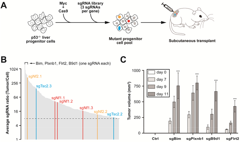
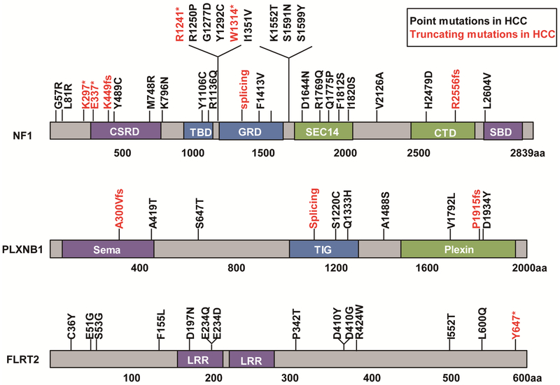
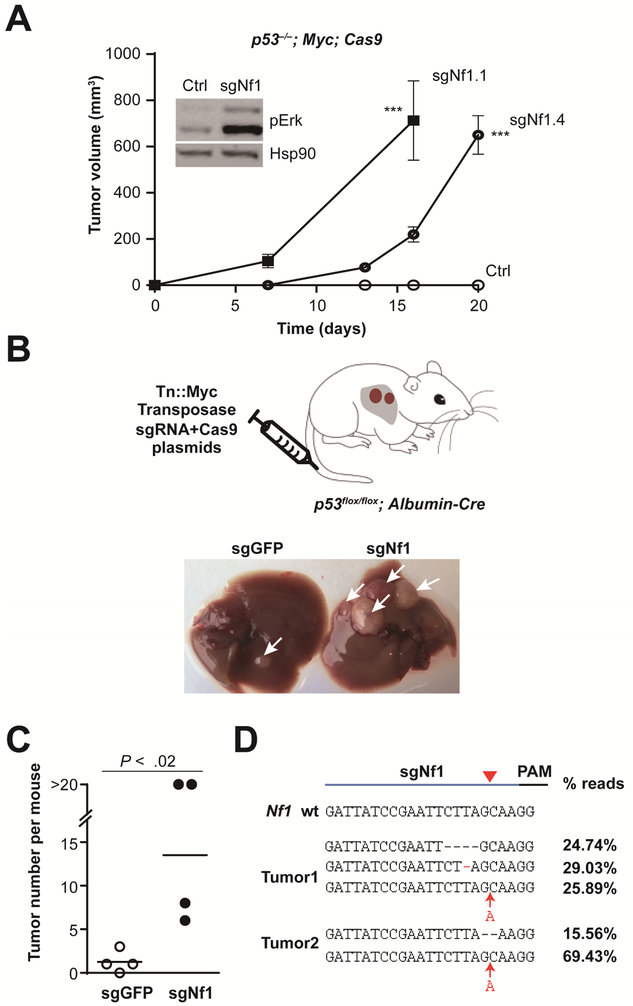
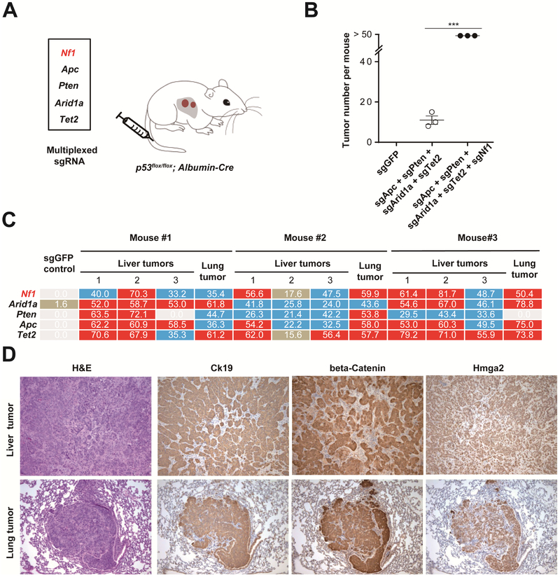
Methods: We performed a genome-wide CRISPR/Cas9-based knockout screen of P53-null mouse embryonic liver progenitor cells that overexpressed MYC. We infected p53-/-;Myc;Cas9 hepatocytes with the mGeCKOa lentiviral library of 67,000 single-guide RNAs (sgRNAs), targeting 20,611 mouse genes, and transplanted the transduced cells subcutaneously into nude mice. Within 1 month, all the mice that received the sgRNA library developed subcutaneous tumors. We performed high-throughput sequencing of tumor DNA and identified sgRNAs increased at least 8-fold compared to the initial cell pool. To validate the top 10 candidate tumor suppressors from this screen, we collected data from patients with hepatocellular carcinoma (HCC) using the Cancer Genome Atlas and COSMIC databases. We used CRISPR to inactivate candidate tumor suppressor genes in p53-/-;Myc;Cas9 cells and transplanted them subcutaneously into nude mice; tumor formation was monitored and tumors were analyzed by histology and immunohistochemistry. Mice with liver-specific disruption of p53 were given hydrodynamic tail-vein injections of plasmids encoding Myc and sgRNA/Cas9 designed to disrupt candidate tumor suppressors; growth of tumors and metastases was monitored. We compared gene expression profiles of liver cells with vs without tumor suppressor gene disrupted by sgRNA/Cas9. Genes found to be up-regulated after tumor suppressor loss were examined in liver cancer cell lines; their expression was knocked down using small hairpin RNAs, and tumor growth was examined in nude mice. Effects of the MEK inhibitors AZD6244, U0126, and trametinib, or the multi-kinase inhibitor sorafenib, were examined in human and mouse HCC cell lines.

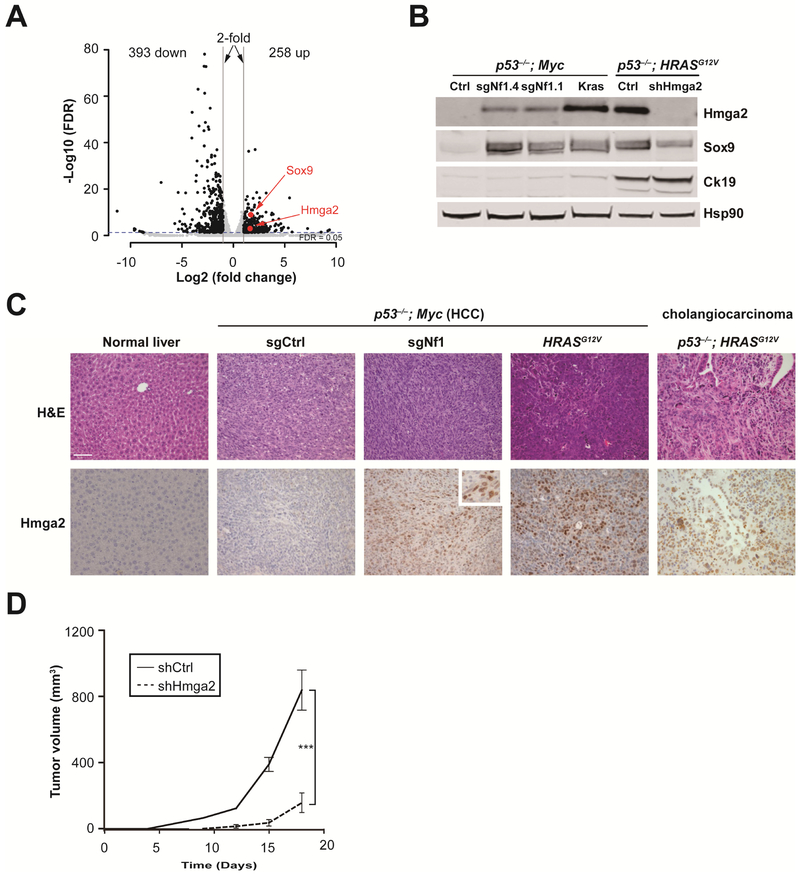
Results: We identified 4 candidate liver tumor suppressor genes not previously associated with liver cancer (Nf1, Plxnb1, Flrt2, and B9d1). CRISPR-mediated knockout of Nf1, a negative regulator of RAS, accelerated liver tumor formation in mice. Loss of Nf1 or activation of RAS up-regulated the liver progenitor cell markers HMGA2 and SOX9. RAS pathway inhibitors suppressed the activation of the Hmga2 and Sox9 genes that resulted from loss of Nf1 or oncogenic activation of RAS. Knockdown of HMGA2 delayed formation of xenograft tumors from cells that expressed oncogenic RAS. In human HCCs, low levels of NF1 messenger RNA or high levels of HMGA2 messenger RNA were associated with shorter patient survival time. Liver cancer cells with inactivation of Plxnb1, Flrt2, and B9d1 formed more tumors in mice and had increased levels of mitogen-activated protein kinase phosphorylation.

Conclusions: Using a CRISPR-based strategy, we identified Nf1, Plxnb1, Flrt2, and B9d1 as suppressors of liver tumor formation. We validated the observation that RAS signaling, via mitogen-activated protein kinase, contributes to formation of liver tumors in mice. We associated decreased levels of NF1 and increased levels of its downstream protein HMGA2 with survival times of patients with HCC. Strategies to inhibit or reduce HMGA2 might be developed to treat patients with liver cancer.

Keywords: CRISPR Screen; Liver Cancer; Mouse Model; Tumor Suppressor Genes.


Conflict of interest statement

Conflicts of interest The authors disclose no conflicts.

Figures

Figure 1.
Figure 1.. Genome-wide ex vivo CRISPR screen identifies new liver tumor suppressor genes.
(A) Outline of the screening strategy sgRNAs targeting tumor suppressors accelerate formation of subcutaneous tumors and are enriched in the tumor. (B) Average ratio of 267 individual sgRNAs enriched > 8-fold in tumors versus cell pool measured by high-throughput sequencing (n = 8). All three Nf1 sgRNAs (sgNf1.1, 2, 3) were enriched. Known liver tumor suppressors (Nf2, Tsc2) with two enriched sgRNAs, and new candidates (Bim, Plxnb1, etc) with one enriched sgRNA are highlighted. (C) Validation of a subset of top-scoring sgRNAs in the subcutaneous tumor assay (n = 4 tumors). Average volume (n = 4) of tumors derived p53−/−; Myc; Cas9 cells infected with a control sgGFP (Ctrl) and a subset of top-scoring sgRNAs (sgBim, sgPlxnb1, sgB9d1, sgFlrt2). ***, P < .001. Error bars, mean ± s.e.m.
Figure 2.
Figure 2.. Point mutations in NF1, PLXNB1 and FLRT2 in human HCC.
* denotes nonsense mutation. Fs denotes frameshift mutation. CSRD, a cysteine-serine-rich domain; TBD, a tubulin-binding domain; GRD, a central GTPase-activating protein-related domain; SBD, a syndecan-binding domain; LRR, Leucine-rich repeat. Data are from TCGA and COSMIC.
Figure 3.
Figure 3.. Nf1 is a bona fide liver tumor suppressor.
(A) Two individual Nf1 sgRNAs accelerated subcutaneous tumor growth. The tumors (n = 4) were derived from p53; Myc; Cas9 cells (Ctrl) infected with sgNf1.1 and sgNf1.4, independently. Error bars, mean ± s.e.m. The inset shows that sgNfl unregulated the phosphor-Erk (pErk) with Hsp90 as a loading control. ***, P < .001. Error bars, mean ± s.e.m. (B) Schematic of hydrodynamic delivery of plasmids encoding Myc transposon (Tn) and Cas9/sgRNA (targeting Nf1, or GFP control) into liver-specific p53-knockout mouse. Representative images of livers from mice treated with sgGFP control (left) or sgNf1 (right) at 1 month after injection are shown. Arrows denote tumors. (C) Quantification of tumor number per mouse (n = 4 mice) shown in (B). (D) Sequences of Nf1 sgRNA target sites from representative liver tumors showing indel mutations and the fraction of reads for each.
Figure 4.
Figure 4.. Multiplexed CRISPR accelerates tumor formation.
(A) sgRNA mixture was injected into p53flox/flox; Albumin-Cre mice by hydrodynamic injection. (B) Quantification of tumor number per mouse (n = 3 mice) at 2 months shown in (A). ***, P < .001. Error bars, mean ± s.e.m. (C) Deep sequencing for the representative liver and lung tumors at 3 months showing indel percentages at the assayed target sites (> 50 shown with red background, between 20 and 50 blue background, between 0 and 20 brown background, 0 white background). The varying indel rates may have been caused by different percentages of wild-type stromal cells within each tumor (see panel D). One liver tumor and one lung tumor had a low percentage of Pten indels. (D) H&E and IHC. All lung tumors show Ck19 staining (10 × lens).
Figure 5.
Figure 5.. Nf1 negatively regulates Ras-dependent activation of liver progenitor cell markers Hmga2 and Sox9.
(A) Volcano plot of mRNA levels in Nf1 sgRNA-treated cells compared to the control sgRNA-treated cells (n = 3). (B) Western blot shows increased Hmga2 and Sox9 levels upon sgRNA-inactivation of Nf1 or activation of Kras (KrasG12D cDNA) in p53−/−; Myc liver cells. Hmga2, Sox9 and Ck19 levels were high in p53−/−; HRASG12V cholangiocarcinoma cells. (C) Immunohistochemistry of representative liver sections showing increased Hmga2 protein upon sgRNA-inactivation of Nf1 or activation of HRASG12V in p53−/−; Myc tumors, and in the p53−/−; HRASG12V cholangiocarcinoma (CCA) mouse model (n ≥ 3 mice per group). Normal liver serves as a control. Scale bar is 50 μm. Inset shows a high-magnification view. (D) Graph of subcutaneous tumor volumes in nude mice receiving p53−/−; HRASG12V cells treated with Hmga2 shRNA (shHmga2) or control shRNA (shCtrl). ***, P < .001. Error bars are s.d. of mean (n = 4 tumors).
Figure 6.
Figure 6.. Sorafenib or MEK inhibitors can suppress HMGA2 and SOX9 expression in liver cancer cell lines.
(A) and (C) qPCR showing the levels of HMGA2 and SOX9 mRNA in Hep3B and Huh7 human HCC cells treated for 48 hours with 1 μM AZD6244, or 7.5 μM sorafenib relative to control-treated cells (Ctrl). (B) and (D) qPCR showing Hmga2 and Sox9 expression in p53−/−; HRASG12V and p53−/−; Myc; Cas9; sgNf1 mouse liver cells. Drug treatment was the same as in (A). Error bars are s.d. of mean (n = 3). ***, P < .001. (E) and (F) Trametinib, a MEK inhibitor, suppressed colony formation in both human and mouse liver cancer cell lines.
Figure 7.
Figure 7.. NF1 and HMGA2 correlate with patient survival in liver cancer.
(A-B) Survival curves of patients expressing high (Top 40%) or low (Bottom 40%) levels of NF1 mRNA (A) or HMGA2 mRNA (B). Based on data from a clinical study . (C) Summary of NF1 and other selected RAS pathway mutations in TCGA liver cancer patients (n = 373). Each column (light grey bars) represents one patient. Dark grey denotes patients with mutations. RASA1 (p120 RAS GAP) and RP6SKA3 are inhibitors of RAS pathway. RP6SKA3 mutations have been reported in HCC . (D) Schematic that loss of Nf1, Plxnb1, Flrt2 or B9d1 convergently activates the MAPK pathway and induces liver progenitor cell markers HMGA2/SOX9 in liver cancer. Green and red arrows denote decreased or increased activity.

Comment in

References

    1. Zucman-Rossi J, Villanueva A, Nault JC, et al. Genetic landscape and biomarkers of hepatocellular carcinoma. Gastroenterology 2015;149:1226–1239. - PubMed
    1. Farazi PA, DePinho RA. Hepatocellular carcinoma pathogenesis: from genes to environment. Nat Rev Cancer 2006;6:674–87. - PubMed
    1. Marquardt JU, Andersen JB, Thorgeirsson SS. Functional and genetic deconstruction of the cellular origin in liver cancer. Nat Rev Cancer 2015;15:653–67. - PubMed
    1. Torre LA, Bray F, Siegel RL, et al. Global cancer statistics, 2012. CA: A Cancer Journal for Clinicians 2015;65:87–108. - PubMed
    1. Llovet JM, Ricci S, Mazzaferro V, et al. Sorafenib in advanced hepatocellular carcinoma. N Engl J Med 2008;359:378–90. - PubMed

MeSH terms

Substances