. 2017 Apr 1:52:21-32.
doi: 10.1016/j.actbio.2016.12.023. Epub 2016 Dec 10.

Defining the hierarchical organisation of collagen VI microfibrils at nanometre to micrometre length scales

Affiliations

Defining the hierarchical organisation of collagen VI microfibrils at nanometre to micrometre length scales

Alan R F Godwin et al. Acta Biomater. .

Abstract

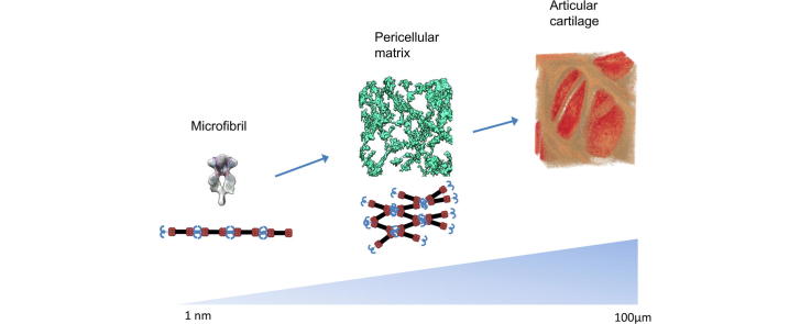
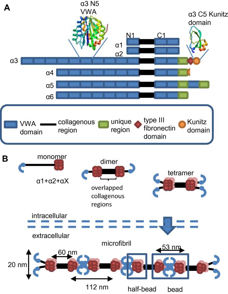
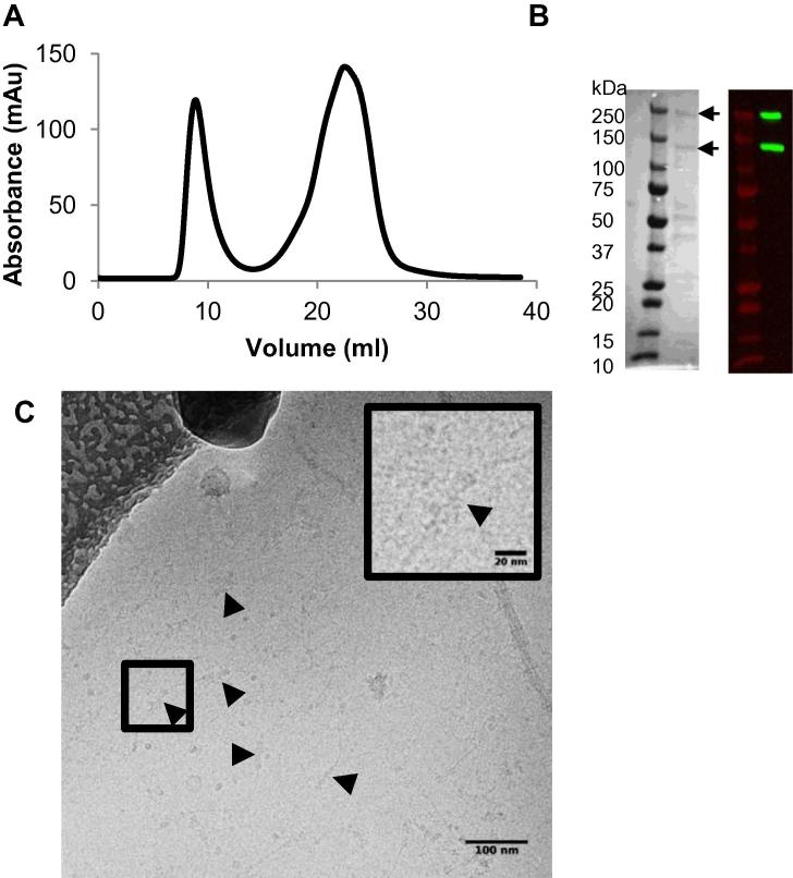
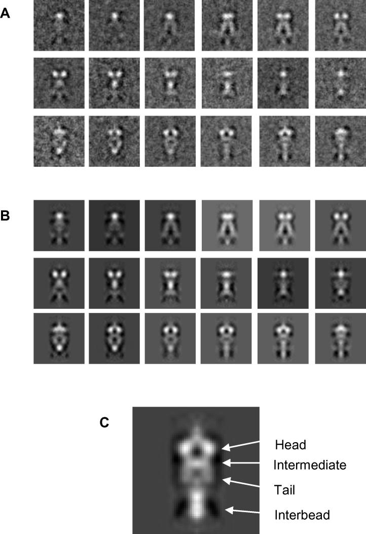
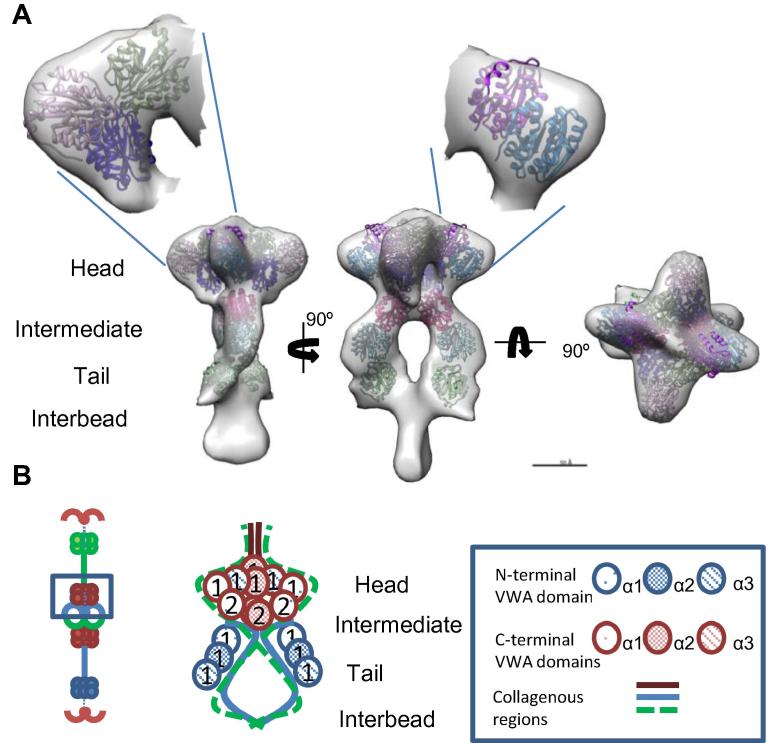
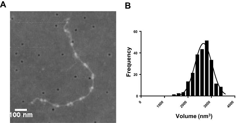
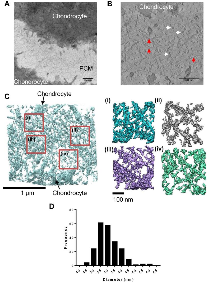
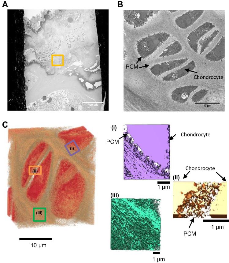
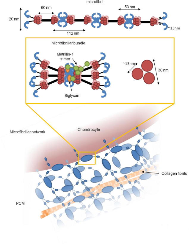
Extracellular matrix microfibrils are critical components of connective tissues with a wide range of mechanical and cellular signalling functions. Collagen VI is a heteromeric network-forming collagen which is expressed in tissues such as skin, lung, blood vessels and articular cartilage where it anchors cells into the matrix allowing for transduction of biochemical and mechanical signals. It is not understood how collagen VI is arranged into microfibrils or how these microfibrils are arranged into tissues. Therefore we have characterised the hierarchical organisation of collagen VI across multiple length scales. The frozen hydrated nanostructure of purified collagen VI microfibrils was reconstructed using cryo-TEM. The bead region has a compact hollow head and flexible tail regions linked by the collagenous interbead region. Serial block face SEM imaging coupled with electron tomography of the pericellular matrix (PCM) of murine articular cartilage revealed that the PCM has a meshwork-like organisation formed from globular densities ∼30nm in diameter. These approaches can characterise structures spanning nanometer to millimeter length scales to define the nanostructure of individual collagen VI microfibrils and the micro-structural organisation of these fibrils within tissues to help in the future design of better mimetics for tissue engineering.

Statement of significance: Cartilage is a connective tissue rich in extracellular matrix molecules and is tough and compressive to cushion the bones of joints. However, in adults cartilage is poorly repaired after injury and so this is an important target for tissue engineering. Many connective tissues contain collagen VI, which forms microfibrils and networks but we understand very little about these assemblies or the tissue structures they form. Therefore, we have use complementary imaging techniques to image collagen VI microfibrils from the nano-scale to the micro-scale in order to understand the structure and the assemblies it forms. These findings will help to inform the future design of scaffolds to mimic connective tissues in regenerative medicine applications.

Keywords: Articular cartilage; Collagen VI; Pericellular matrix; SBF-SEM; Tomography.


Figures

None
Graphical abstract
Fig. 1
Fig. 1
Domain organisation of collagen VI α-chains and microfibril formation. A) A cartoon illustrating the domain organisation of the collagen VI α chains. The VWA domains are numbered from N1 and C1 from the closest domains to the collagenous region. Also shown are cartoon representations of the structures of the α3 chain N5 VWA domain and the α3 chain Kunitz domain . The domain cartoons are rainbow coloured from blue at the N-terminus to red at the C-terminus. B) A cartoon representation of collagen VI microfibril assembly. Collagen VI heteromeric monomers form from an α1, α2 and αX chain where X can be α-chains 3–6. Triple-helical monomers then form disulphide linked dimers and then tetramers before being secreted into the extracellular space where microfibrils are formed. C-terminal globular regions are shown in red, N-terminal regions are shown in blue. The bead and half-bead regions of the microfibril are highlighted. The bead region contains the same number of VWA domains as a tetramer with the half-bead being equivalent to a dimer. The mature microfibril contains 10 C-terminal VWA domains in each half-bead . (For interpretation of the references to colour in this figure legend, the reader is referred to the web version of this article.)
Fig. 2
Fig. 2
Purification and imaging of collagen VI microfibrils from bovine cornea. (A) Size exclusion chromatography of collagenase extracted bovine corneal tissue using a Sepharose Cl-2B column. The absorbance (mAu) at 280 nm is plotted against the elution volume (ml). The first peak represents the void volume of the column where microfibrils elute. (B) Reducing SDS-PAGE (left hand panel) and western blot (right hand panel) of the central fraction of the void peak. Collagen VI chains were detected using a polyclonal rabbit anti collagen VI antibody. Arrows highlight bands at approximately 250 kDa, which corresponds to the α3 chain, and at 120 kDa which corresponds to α1 and α2 chains. C) Bovine collagen VI was imaged under cryo conditions using a FEI Tecnai G2 Polara TEM operating at an accelerating voltage of 200KV. Black arrows highlight the globular bead regions. A magnified image of a bead region is shown in the top right of the figure.
Fig. 3
Fig. 3
3D reconstruction of the collagen VI microfibril bead region. Individual images of collagen VI bead regions were cropped from cryo-TEM images using EMAN2 and aligned using FindEM . Half-bead particles were extracted from aligned stacks of beads before being reconstructed into a 3D model using single particle reconstruction methods using FindEM and SPIDER . (A) Class-sum images of aligned particles. Particles were classified by similarity to model projections using cross-correlation. (B) Reprojections of the final half-bead model. Class-sum images and model reprojections represent 10° rotations around the collagen VI fibre axis. (C) The central slice from a radial average of the collagen VI half-bead model. The box size is 77 × 77 nm for all panels.
Fig. 4
Fig. 4
3D structure of collagen VI microfibril. (A) Cryo-TEM structure of collagen VI visualised using UCSF Chimera . VWA domains were placed in the electron density map by hand before their fit was optimised using the UCSF Chimera fit in map tool . Ten C-terminal VWA domains were fitted into the head region and 3 VWA domains were fit in the intermediate and tail regions. (B) Schematic model of the organisation of VWA domains in the half-bead structure. Three VWA domains were fitted in each of the larger lobe-like structures, potentially corresponding to the VWA C1 and C2 from either α1 or α2 chain and C1 from the α3 chain, and each of the smaller lobes could accommodate the remaining two VWA domains from either the α1 or the α2 chain. The intermediate and tail region could accommodate three VWA domains which could correspond to the N1 VWA domains from the α1, 2 and 3 chains.
Fig. 5
Fig. 5
AFM analysis of collagen VI microfibrils. (A) An AFM image of isolated collagen VI microfibrils adsorbed onto a glass cover slip. (B) A histogram of collagen VI bead region volumes. A Gaussian curve was fitted to the data using non-linear regression in GraphPad Prism version 6.04. The data fit with an R square of 0.972 and had a calculated mean value of 2662 nm3. A total of 225 bead regions were measured.
Fig. 6
Fig. 6
Murine articular cartilage electron tomography. Articular cartilage was imaged using TEM and a tilt series collected. (A) Representative image of the PCM between two chondrocytes. (B) A virtual z-slice from a tomogram of the chondrocyte PCM. Highlighted with white arrows are globular densities which are potentially bundles of collagen VI and red arrows indicate straighter fibrillar structures potentially collagen II fibrils (C) A tomogram rendered using UCSF Chimera. Red boxes define regions of interest magnified in panels i–iv. (D) Diameters of PCM globular densities were measured using ImageJ and plotted as a histogram of their diameters. The mean diameter was 30.4 nm ± 0.5 nm (SEM). A total of 218 particles were measured from one tomogram. (For interpretation of the references to colour in this figure legend, the reader is referred to the web version of this article.)
Fig. 7
Fig. 7
Murine articular cartilage imaged using SBF-SEM. Murine articular cartilage tissue was imaged using SBF-SEM. (A) An image of the sample block face is shown, highlighted is the region where the SBF-SEM data set was collected. (B) A representative image from the SBF-SEM data set where the PCM and chondrocyte are labelled. (C) 3D reconstruction of a sub-volume of the SBF-SEM data-set (dimensions of 27 × 23 × 16 μm), the right panel shows 3D reconstructions of the PCM surrounding chondrocytes (i and ii) and territorial matrix (iii).
Fig. 8
Fig. 8
Model of collagen VI hierarchical organisation A collagen VI microfibril forms bundles potentially through interaction with adaptor complexes such as biglycan via the collagen VI N-terminal regions . Shown in the right hand panel is a schematic diagram of a cross-section of a bundle of three microfibrils which are ∼13 nm in diameter, forming a complex ∼30 nm in diameter. Microfibrillar bundles can then form larger hexagonal networks in the PCM. The microfibrillar bundles become nodes which are connected by individual microfibrils.

References

    1. Hunziker E.B., Quinn T.M., Hauselmann H.J. Quantitative structural organization of normal adult human articular cartilage. Osteoarthritis Cartilage/OARS. 2002;10(7):564–572. Osteoarthritis Research Society. - PubMed
    1. Correa D., Lietman S.A. Articular cartilage repair: Current needs, methods and research directions. Semin. cell Dev. Biol. 2016 - PubMed
    1. Guilak F., Estes B.T., Diekman B.O., Moutos F.T., Gimble J.M. 2010 Nicolas Andry Award: multipotent adult stem cells from adipose tissue for musculoskeletal tissue engineering. Clin. Orthop. Relat. Res. 2010;468(9):2530–2540. - PMC - PubMed
    1. Awad H.A., Wickham M.Q., Leddy H.A., Gimble J.M., Guilak F. Chondrogenic differentiation of adipose-derived adult stem cells in agarose, alginate, and gelatin scaffolds. Biomaterials. 2004;25(16):3211–3222. - PubMed
    1. Zelenski N.A., Leddy H.A., Sanchez-Adams J., Zhang J., Bonaldo P., Liedtke W., Guilak F. Type VI collagen regulates pericellular matrix properties, chondrocyte swelling, and mechanotransduction in mouse articular cartilage. Arthritis Rheumatol. 2015;67(5):1286–1294. (Hoboken, N.J.) - PMC - PubMed

Publication types

MeSH terms

Substances

LinkOut - more resources