Silent Cerebral Events after Atrial Fibrillation Ablation - Overview and Current Data
- PMID: 27957043
- PMCID: PMC4956131
- DOI: 10.4022/jafib.996
Silent Cerebral Events after Atrial Fibrillation Ablation - Overview and Current Data
Abstract
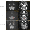
Silent cerebral lesions (SCL) have been identified on brain magnetic resonance imaging (MRI) in apparently asymptomatic patients after cardiovascular procedures. After atrial fibrillation (AF) ablation incidences range from 1 to over 40% depending upon different factors. MRI definition should include diffusion weighted imaging (DWI) to detect hyperintensities (bright spots) due to acute brain ischemia correlated with a hypointensity in the apparent diffusion coefficient mapping (ADC-map) to rule out artifacts. The genesis of SCL appears to be multifactorial and appears to be a result of embolic events either from gaseous or solid particles. The MRI pattern appears to be comparable not hinting towards a specific mechanism. One may distinguish two different MRI definition: one, more sensitive, for silent ischemic events (SCE) not proven to be related to cell death (DWI positive but FLAIR negative); and one for SCL that are due to edema caused by cell death which will lead to glial cell scar formation (DWI positive and FLAIR positive). For ease of data interpretation, future studies should ensure both definitions, and that DWI and FLAIR data is acquired using identical slice thickness and orientation. Risk factors associated with increased SCL-incidences involve patient-specific, technology-associated and procedural determinants. When using a high-sensitive MRI definition differences in SCE-rates in between technologies appear to be less prominent. Further studies on the effects of different periprocedural anticoagulation regimen, different steps of the ablation procedure and new technologies are needed. For now, SCL incidence may determine the thrombogenic potential of an ablation technology and further studies to reduce or avoid SCL generation are desirable. It appears reasonable, that any SCE should be avoided.
Figures


References
-
- Schrickel Jan Wilko, Lickfett Lars, Lewalter Thorsten, Mittman-Braun Erica, Selbach Stephanie, Strach Katharina, Nähle Claas P, Schwab Jörg Otto, Linhart Markus, Andrié Rene, Nickenig Georg, Sommer Torsten. Incidence and predictors of silent cerebral embolism during pulmonary vein catheter ablation for atrial fibrillation. Europace. 2010 Jan;12 (1):52–7. - PubMed
-
- Rodés-Cabau Josep, Dumont Eric, Boone Robert H, Larose Eric, Bagur Rodrigo, Gurvitch Ronen, Bédard Fernand, Doyle Daniel, De Larochellière Robert, Jayasuria Cleonie, Villeneuve Jacques, Marrero Alier, Côté Mélanie, Pibarot Philippe, Webb John G. Cerebral embolism following transcatheter aortic valve implantation: comparison of transfemoral and transapical approaches. J. Am. Coll. Cardiol. 2011 Jan 4;57 (1):18–28. - PubMed
-
- Wieczorek Marcus, Hoeltgen Reinhard, Brueck Martin. Does the number of simultaneously activated electrodes during phased RF multielectrode ablation of atrial fibrillation influence the incidence of silent cerebral microembolism? Heart Rhythm. 2013 Jul;10 (7):953–9. - PubMed
-
- Jurga Juliane, Nyman Jesper, Tornvall Per, Mannila Maria Nastase, Svenarud Peter, van der Linden Jan, Sarkar Nondita. Cerebral microembolism during coronary angiography: a randomized comparison between femoral and radial arterial access. Stroke. 2011 May;42 (5):1475–7. - PubMed
-
- Rillig Andreas, Meyerfeldt Udo, Tilz Roland Richard, Talazko Jochen, Arya Anita, Zvereva Vlada, Birkemeyer Ralf, Miljak Tomislav, Hajredini Bajram, Wohlmuth Peter, Fink Ulrich, Jung Werner. Incidence and long-term follow-up of silent cerebral lesions after pulmonary vein isolation using a remote robotic navigation system as compared with manual ablation. Circ Arrhythm Electrophysiol. 2012 Feb;5 (1):15–21. - PubMed
Publication types
LinkOut - more resources
Full Text Sources
Research Materials
Miscellaneous
