

Comparison of Two Base Materials Regarding Their Effect on Root Canal Treatment Success in Primary Molars with Furcation Lesions
- PMID: 27957486
- PMCID: PMC5121461
- DOI: 10.1155/2016/1429286
Comparison of Two Base Materials Regarding Their Effect on Root Canal Treatment Success in Primary Molars with Furcation Lesions
Abstract
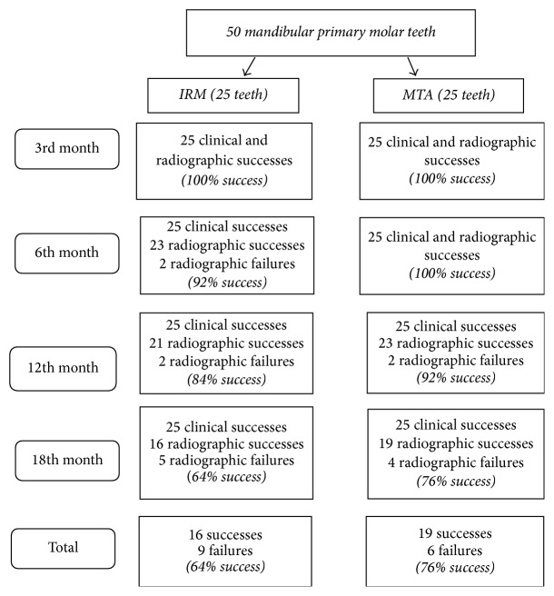
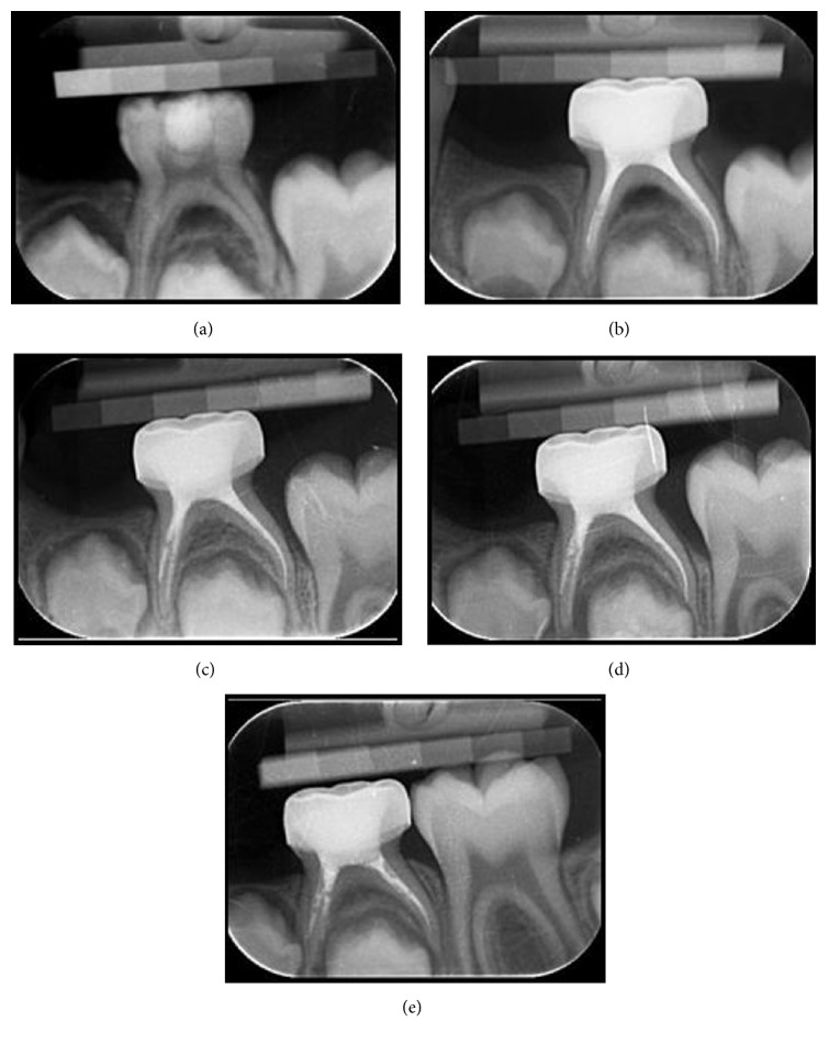
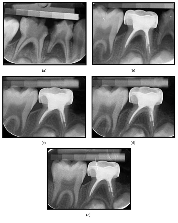
Introduction. The aim of this study was to compare MTA with another base material, IRM, which is generally used on pulpal floor after root canal treatment, regarding their effect on the success of root canal treatment of primary teeth with furcation lesions. Materials and Methods. Fifty primary teeth with furcation lesions were divided into 2 groups. Following root canal treatment, the pulpal floor was coated with MTA in the experimental group and with IRM in the control group. Teeth were followed up considering clinical (pain, pathological mobility, tenderness to percussion and palpation, and any soft tissue pathology and sinus tract) and radiographical (pathological root resorption, reduced size or healing of existing lesion, and absence of new lesions at the interradicular or periapical area) criteria for 18 months. For the statistical analysis, Fisher's exact test and Pearson's chi-square tests were used and a p value of <0.05 was considered to be statistically significant. Results. Although there were no statistically significant differences between two groups in terms of treatment success, lesions healed significantly faster in the MTA group. Conclusion. In primary teeth with furcation lesions, usage of MTA on the pulpal floor following root canal treatment can be a better alternative since it induced faster healing.
Conflict of interest statement
The authors declare that they have no competing interests.
Figures
References
-
- Camp J. H., Fuks A. B. Pediatric endodontics: endodontic treatment for the primary and young permanent dentition. In: Cohen S., Hargreaves K. M., editors. Pathways of the Pulp. 9th. chapter 22. St. Louis, Mo, USA: Mosby; 2006.
-
- Kramer P. F., Faraco Júnior I. M., Meira R. A SEM investigation of accessory foramina in the furcation areas of primary molars. The Journal of Clinical Pediatric Dentistry. 2003;27(2):157–161. - PubMed
-
- Guglielmi C. A. B., Müller Ramalho K., Scaramucci T., Da Silva S. R. E. P., Imparato J. C. P., Pinheiro S. L. Evaluation of the furcation area permeability of deciduous molars treated by neodymium:yttrium-aluminum-garnet laser or adhesive. Lasers in Medical Science. 2010;25(6):873–880. doi: 10.1007/s10103-009-0730-z. - DOI - PubMed
-
- Dammaschke T., Witt M., Ott K., Schäfer E. Scanning electron microscopic investigation of incidence, location, and size of accessory foramina in primary and permanent molars. Quintessence International. 2004;35(9):699–705. - PubMed
Publication types
MeSH terms
Substances
LinkOut - more resources
Full Text Sources
Other Literature Sources
