. 2016 Dec 5;4(1):e307.
doi: 10.1212/NXI.0000000000000307. eCollection 2017 Jan.

Neurochondrin is a neuronal target antigen in autoimmune cerebellar degeneration

Affiliations

Neurochondrin is a neuronal target antigen in autoimmune cerebellar degeneration

Ramona Miske et al. Neurol Neuroimmunol Neuroinflamm. .

Abstract

Objective: To report on a novel neuronal target antigen in 3 patients with autoimmune cerebellar degeneration.

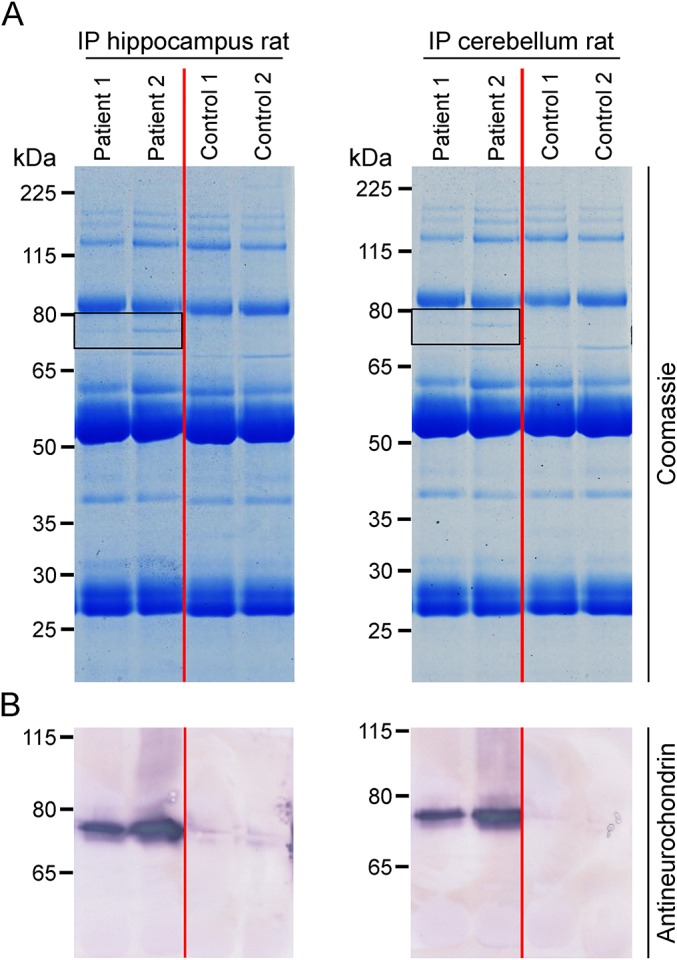
Methods: Three patients with subacute to chronic cerebellar ataxia and controls underwent detailed clinical and neuropsychological assessment together with quantitative high-resolution structural MRI. Sera and CSF were subjected to comprehensive autoantibody screening by indirect immunofluorescence assay (IFA) and immunoblot. Immunoprecipitation with lysates of hippocampus and cerebellum combined with mass spectrometric analysis was used to identify the autoantigen, which was verified by recombinant expression in HEK293 cells and use in several immunoassays. Multiparameter flow cytometry was performed on peripheral blood and CSF, and peripheral blood was subjected to T-cell receptor spectratyping.

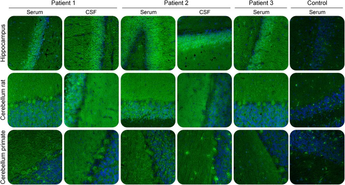
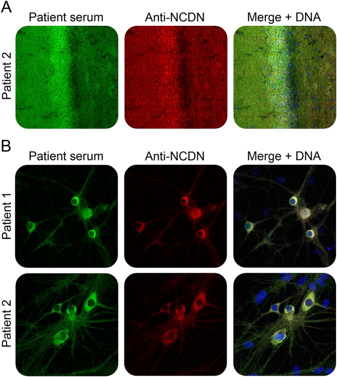
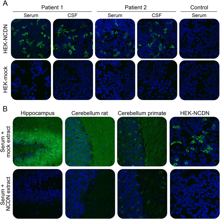
Results: Patients presented with a subacute to chronic cerebellar and brainstem syndrome. MRI was consistent with cortical and cerebellar gray matter atrophy associated with subsequent neuroaxonal degeneration. IFA screening revealed strong immunoglobulin G1 reactivity in sera and CSF with hippocampal and cerebellar molecular and granular layers, but not with a panel of 30 recombinantly expressed established neural autoantigens. Neurochondrin was subsequently identified as the target antigen, verified by IFA and immunoblot with HEK293 cells expressing human neurochondrin as well as the ability of recombinant neurochondrin to neutralize the autoantibodies' tissue reaction. Immune phenotyping revealed intrathecal accumulation and activation of B and T cells during the acute but not chronic phase of the disease. T-cell receptor spectratyping suggested an antigen-specific T-cell response accompanying the formation of antineurochondrin autoantibodies. No such neurochondrin reactivity was found in control cohorts of various neural autoantibody-associated neurologic syndromes, relapsing-remitting multiple sclerosis, cerebellar type of multiple system atrophy, hereditary cerebellar ataxias, other neurologic disorders, or healthy donors.

Conclusion: Neurochondrin is a neuronal target antigen in autoimmune cerebellar degeneration.


Figures

Figure 1
Figure 1. Immunofluorescence staining of central nervous tissues
Cryosections were incubated with patient or control sera (1:100) or CSF (undiluted) in the first step, and with Alexa488-labelled goat anti-human immunoglobulin G in the second step. Nuclei were counterstained by incubation with TO-PRO-3 iodide (blue). A fine granular staining of both granular and molecular layers was obtained with the strongest reaction on the granular cell layer of rat hippocampus. ×200 magnification.
Figure 2
Figure 2. Immunoprecipitation and antigen identification
Lysates of rat hippocampus and cerebellum were incubated with patient or control sera (1:33). Immunocomplexes were isolated with protein-G-coated magnetic beads, eluted by sodium dodecyl sulfate, and subjected to sodium dodecyl sulfate polyacrylamide gel electrophoresis analysis followed by (A) staining with colloidal Coomassie or (B) Western blot using polyclonal rabbit antineurochondrin. Frames indicate the position of the immunoprecipitated antigen at about 75 kDa.
Figure 3
Figure 3. Double-staining of hippocampal tissue and cells with patient serum and rabbit antineurochondrin antibody
Immunofluorescence staining of rat hippocampus tissue section (A) or formalin-fixed and TritonX-100 permeabilized rat hippocampal neurons (B) with patient sera (green) and antineurochondrin antibody (red). Nuclei were counterstained by incubation with TO-PRO-3 iodide (blue). (A, B) ×200 magnification.
Figure 4
Figure 4. Verification of neurochondrin as the novel autoantigen by indirect immunofluorescence
(A) Indirect immunofluorescence using acetone-fixed neurochondrin or mock-transfected HEK293 cells incubated with 1:1,000 diluted serum or 1:10 diluted CSF of patient 1, patient 2, or a healthy control (green). (B) Neutralization of immunofluorescence reaction on neuronal tissues. Patient serum (green) was preincubated with extracts of HEK293 cells transfected with neurochondrin or with empty vector as control. The extract containing neurochondrin abolished the immune reaction. Nuclei were counterstained by incubation with TO-PRO-3 iodide (blue). (A) Hippocampus tissue section: ×200 magnification. (B) Rat hippocampal neurons: ×400 magnification.

References

    1. Melzer N, Meuth SG, Wiendl H. Neuron-directed autoimmunity in the central nervous system: entities, mechanisms, diagnostic clues, and therapeutic options. Curr Opin Neurol 2012;25:341–348. - PubMed
    1. Lennon VA, Kryzer TJ, Pittock SJ, Verkman AS, Hinson SR. IgG marker of optic-spinal multiple sclerosis binds to the aquaporin-4 water channel. J Exp Med 2005;202:473–477. - PMC - PubMed
    1. Dalmau J, Gleichman AJ, Hughes EG, et al. . Anti-NMDA-receptor encephalitis: case series and analysis of the effects of antibodies. Lancet Neurol 2008;7:1091–1098. - PMC - PubMed
    1. Lai M, Hughes EG, Peng X, et al. . AMPA receptor antibodies in limbic encephalitis alter synaptic receptor location. Ann Neurol 2009;65:424–434. - PMC - PubMed
    1. Lancaster E, Lai M, Peng X, et al. . Antibodies to the GABA(B) receptor in limbic encephalitis with seizures: case series and characterisation of the antigen. Lancet Neurol 2010;9:67–76. - PMC - PubMed