. 2016 Dec 7;2(12):e1601941.
doi: 10.1126/sciadv.1601941. eCollection 2016 Dec.

A single splice site mutation in human-specific ARHGAP11B causes basal progenitor amplification

Affiliations

A single splice site mutation in human-specific ARHGAP11B causes basal progenitor amplification

Marta Florio et al. Sci Adv. .

Abstract

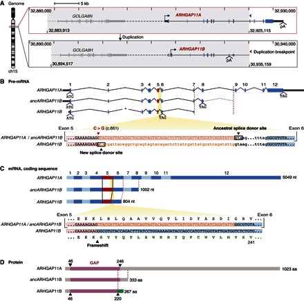
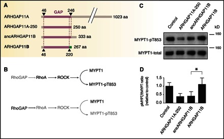
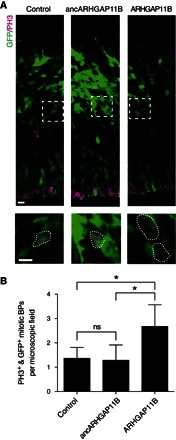
The gene ARHGAP11B promotes basal progenitor amplification and is implicated in neocortex expansion. It arose on the human evolutionary lineage by partial duplication of ARHGAP11A, which encodes a Rho guanosine triphosphatase-activating protein (RhoGAP). However, a lack of 55 nucleotides in ARHGAP11B mRNA leads to loss of RhoGAP activity by GAP domain truncation and addition of a human-specific carboxy-terminal amino acid sequence. We show that these 55 nucleotides are deleted by mRNA splicing due to a single C→G substitution that creates a novel splice donor site. We reconstructed an ancestral ARHGAP11B complementary DNA without this substitution. Ancestral ARHGAP11B exhibits RhoGAP activity but has no ability to increase basal progenitors during neocortex development. Hence, a single nucleotide substitution underlies the specific properties of ARHGAP11B that likely contributed to the evolutionary expansion of the human neocortex.

Keywords: ARHGAP11B; RhoGAP; basal progenitors; brain size; gene duplication; human evolution; human-specific gene; neocortex; neocortical expansion; neurogenesis.


Figures

Fig. 1
Fig. 1. ARHGAP11A and ARHGAP11B genomic, pre-mRNA, mRNA, and protein structures.
(A) Gene structure and genomic context of human ARHGAP11A (top) and ARHGAP11B (bottom). Gray areas indicate the duplicated genomic region (40.642 Mb), which comprises the GOLGA8 and ARHGAP11 genes. Tick marks and numbers indicate genomic coordinates on chromosome 15 (GRCh37/hg19). Red arrowheads point to the 3′ duplication break point, highlighting the partial nature of the ARHGAP11 duplication. Continuous and dashed horizontal lines indicate intragenic and intergenic regions, respectively. Transcription start sites (forward arrows) and polyadenylation sites (pA) are shown. Rectangles indicate exons; dark gray, untranslated regions; blue and red, protein-coding sequences (red, exon 5); white, exonic sequences of ARHGAP11A that are duplicated but typically untranscribed in modern ARHGAP11B. The duplicated GOLGA8 gene 5′ to ARHGAP11 is depicted in light gray. Image was adapted from the University of California Santa Cruz (UCSC) Genome Browser. (B) ARHGAP11A, predicted ancestral ARHGAP11B (ancARHGAP11B), and modern ARHGAP11B pre-mRNAs (top). Translation start (ATG) and stop (TAG) codons are indicated. Numbers (1 to 12) indicate ARHGAP11A exons, 1 to 8 of which were duplicated. Left red dashed line indicates the position of the pA site in exon 7 of modern ARHGAP11B relative to exon 7 of ARHGAP11A and ancARHGAP11B. Dotted intron line 3′ to the predicted ancARHGAP11B stop codon indicates the duplicated portion of ARHGAP11A intron 8 until the duplication break point (right red dashed line). Asterisk indicates a 593-bp deletion in modern ARHGAP11B intron 2. (B) Alignment of ARHGAP11A/B homologous sequences (bottom) encompassing the 3′ end of exon 5 (red background) and 5′ end of exon 6 (blue background), interspaced by intron 5. Exonic and intronic nucleotide (nt) sequences are displayed in uppercase and lowercase letters, respectively. The C→G base substitution (red, position c.661) produces a new splice donor site in modern ARHGAP11B, 55-nt 5′ to the ancestral one. As a consequence, these 55 nt (orange sequences) become intronic. (C) ARHGAP11A, ancARHGAP11B, and modern ARHGAP11B coding sequences (top). Alternating light-dark blue rectangles indicate exons. Black lines highlight the shortening of modern ARHGAP11B exon 5. (C) Alignment of ARHGAP11A/B coding sequences (bottom) encompassing the 3′ end of exon 5 and the 5′ end of exon 6, with the corresponding amino acid sequences (until residue 241) depicted above and below. The 55-nt sequence corresponding to the 3′ end of ARHGAP11A exon 5 (orange) is spliced out from the modern ARHGAP11B mRNA. The resulting frameshift generates a novel C-terminal amino acid sequence (green) unique to modern ARHGAP11B. (B and C) Note that all RNA sequences are depicted with T instead of U. (D) ARHGAP11A, ancARHGAP11B, and modern ARHGAP11B protein structures showing the conserved portions of the ARHGAP11A GAP domain (purple) and the novel C-terminal domain of modern ARHGAP11B (green) starting at residue 221. aa, amino acids.
Fig. 2
Fig. 2. Ancestral but not modern ARHGAP11B shows RhoGAP activity.
(A) Domain structures of ARHGAP11A, ARHGAP11A-250, ancestral ARHGAP11B (ancARHGAP11B), and modern ARHGAP11B. Purple, GAP domain; green, C-terminal domain unique to ARHGAP11B. (B) RhoGAP activity assay in transfected COS-7 cells based on the lack of phosphorylation of MYPT1 at Thr853 (pT853) by inhibition of Rho-associated protein kinase (ROCK/Rho-kinase). Gray indicates reduced activity/presence. (C) Immunoblots of COS-7 cells transfected with empty vector (control), ARHGAP11A-250, ancARHGAP11B, or modern ARHGAP11B using anti-MYPT1-pT853 (top) or anti-MYPT1 (bottom) antibodies; the position of a 160-kDa marker is indicated. (D) Quantification of immunoblots as shown in (C). The ratio of MYPT1-pT853 (pMYPT) to total MYPT1 (tMYPT) was determined, the mean value obtained for the control was set to 1.0, and the mean values of the other three conditions were expressed relative to this. Data are the means of three independent transfections; error bars, SD; *P < 0.05 (paired t test).
Fig. 3
Fig. 3. Modern but not ancestral ARHGAP11B increases BPs in mouse developing neocortex.
In utero electroporation of pCAGGS–green fluorescent protein (GFP) together with empty vector (control), ancestral ARHGAP11B (ancARHGAP11B), and modern ARHGAP11B expression plasmids in E13.5 mouse neocortex followed by analysis at E14.5. (A) GFP (green) and phosphohistone H3 (PH3, magenta) immunofluorescence labeling targeted and mitotic cells, respectively. Areas in dashed boxes are shown at higher magnification at the bottom. Scale bars, 10 μm. (B) Quantification of targeted mitotic BPs, identified by the presence of both GFP and PH3 immunofluorescence at a basal location. Data are the means of four (control), six (ancARHGAP11B), and five (ARHGAP11B) embryos from three litters, expressed per 100-μm-wide field of cortical wall; error bars, SD; *P < 0.05; ns, not significant [analysis of variance (ANOVA) with Bonferroni’s post hoc test for multiple comparisons].

References

    1. G. F. Striedter, Principles of Brain Evolution (Sinauer Associates Inc., Sunderland, 2005).
    1. Rakic P., Evolution of the neocortex: A perspective from developmental biology. Nat. Rev. Neurosci. 10, 724–735 (2009). - PMC - PubMed
    1. Fietz S. A., Huttner W. B., Cortical progenitor expansion, self-renewal and neurogenesis—A polarized perspective. Curr. Opin. Neurobiol. 21, 23–35 (2011). - PubMed
    1. Lui J. H., Hansen D. V., Kriegstein A. R., Development and evolution of the human neocortex. Cell 146, 18–36 (2011). - PMC - PubMed
    1. Borrell V., Reillo I., Emerging roles of neural stem cells in cerebral cortex development and evolution. Dev. Neurobiol. 72, 955–971 (2012). - PubMed