. 2017 Mar 1;595(5):1607-1618.
doi: 10.1113/JP272893. Epub 2017 Feb 5.

Angiotensin II reduces the surface abundance of KV 1.5 channels in arterial myocytes to stimulate vasoconstriction

Affiliations

Angiotensin II reduces the surface abundance of KV 1.5 channels in arterial myocytes to stimulate vasoconstriction

Michael W Kidd et al. J Physiol. .

Abstract

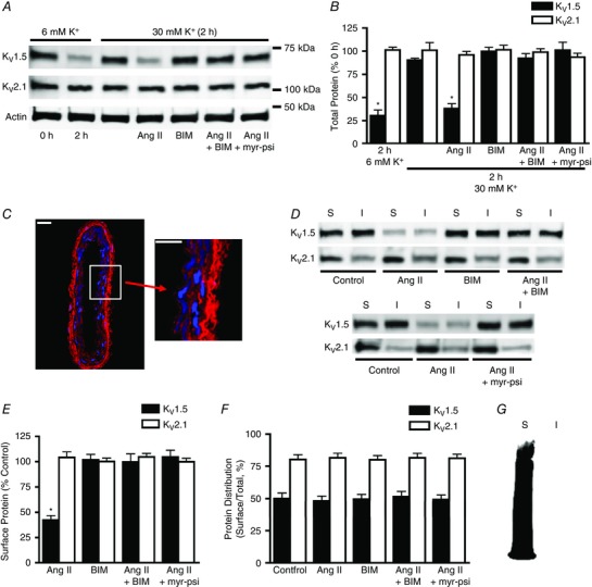
Key points: Several different voltage-dependent K+ (KV ) channel isoforms are expressed in arterial smooth muscle cells (myocytes). Vasoconstrictors inhibit KV currents, but the isoform selectivity and mechanisms involved are unclear. We show that angiotensin II (Ang II), a vasoconstrictor, stimulates degradation of KV 1.5, but not KV 2.1, channels through a protein kinase C- and lysosome-dependent mechanism, reducing abundance at the surface of mesenteric artery myocytes. The Ang II-induced decrease in cell surface KV 1.5 channels reduces whole-cell KV 1.5 currents and attenuates KV 1.5 function in pressurized arteries. We describe a mechanism by which Ang II stimulates protein kinase C-dependent KV 1.5 channel degradation, reducing the abundance of functional channels at the myocyte surface.

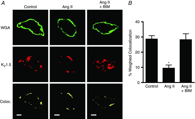
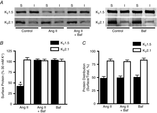
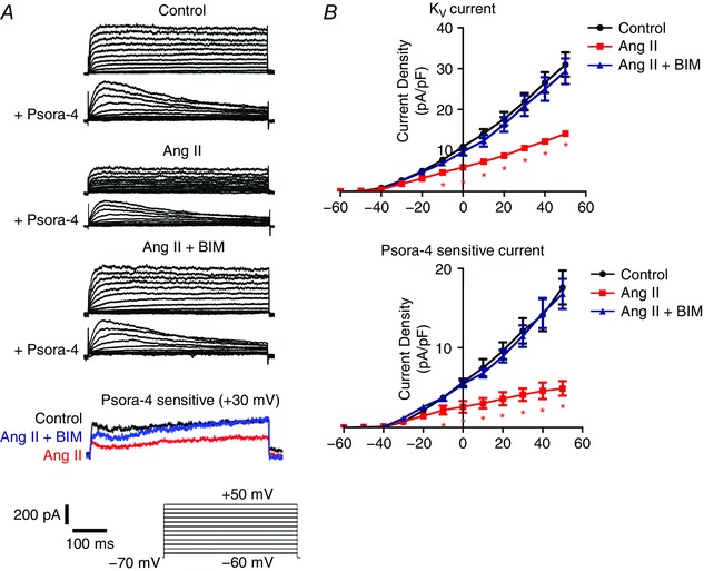
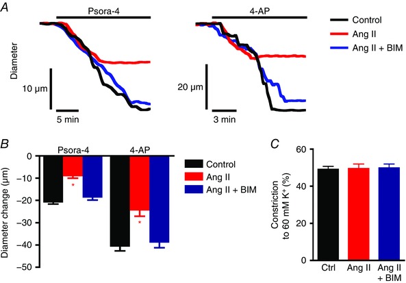
Abstract: Smooth muscle cells (myocytes) of resistance-size arteries express several different voltage-dependent K+ (KV ) channels, including KV 1.5 and KV 2.1, which regulate contractility. Myocyte KV currents are inhibited by vasoconstrictors, including angiotensin II (Ang II), but the mechanisms involved are unclear. Here, we tested the hypothesis that Ang II inhibits KV currents by reducing the plasma membrane abundance of KV channels in myocytes. Angiotensin II (applied for 2 h) reduced surface and total KV 1.5 protein in rat mesenteric arteries. In contrast, Ang II did not alter total or surface KV 2.1, or KV 1.5 or KV 2.1 cellular distribution, measured as the percentage of total protein at the surface. Bisindolylmaleimide (BIM; a protein kinase C blocker), a protein kinase C inhibitory peptide or bafilomycin A (a lysosomal degradation inhibitor) each blocked the Ang II-induced decrease in total and surface KV 1.5. Immunofluorescence also suggested that Ang II reduced surface KV 1.5 protein in isolated myocytes; an effect inhibited by BIM. Arteries were exposed to Ang II or Ang II plus BIM (for 2 h), after which these agents were removed and contractility measurements performed or myocytes isolated for patch-clamp electrophysiology. Angiotensin II reduced both whole-cell KV currents and currents inhibited by Psora-4, a KV 1.5 channel blocker. Angiotensin II also reduced vasoconstriction stimulated by Psora-4 or 4-aminopyridine, another KV channel inhibitor. These data indicate that Ang II activates protein kinase C, which stimulates KV 1.5 channel degradation, leading to a decrease in surface KV 1.5, a reduction in whole-cell KV 1.5 currents and a loss of functional KV 1.5 channels in myocytes of pressurized arteries.

Keywords: KV channel; angiotensin II; smooth muscle; vasoconstriction.


Figures

Figure 1
Figure 1. Angiotensin II reduces total and surface KV1.5 channel protein through protein kinase C (PKC) activation in mesenteric arteries
A, representative Western blot images of total arterial KV1.5, KV2.1 and actin protein at 0 or 2 h in 6 mm K+ physiological saline solution (PSS), 30 mm K+ PSS, or 30 mm K+ PSS with angiotensin II (Ang II; 100 nm), bisindolylmaleimide (BIM; 10 μm), Ang II plus BIM, or a myristoylated PKC inhibitory peptide (myr‐psi; 100 μm). B, mean data. n = 6 for each. * P < 0.05 vs. 0 h. C, immunofluorescence of a section cut from a biotinylated mesenteric artery labelled with Alexa Fluor 546 streptavidin (red) and DAPI (blue), a nuclear stain. Scale bars represent 20 μm. D, representative Western blot images of arterial biotinylation samples illustrating surface (S) and intracellular (I) KV1.5 and KV2.1 following 2 h in 30 mm K+ alone (control) or in the presence of Ang II (100 nm), BIM (10 μm), Ang II plus BIM, or Ang II plus myr‐psi (100 μm). E, mean data for surface protein in conditions from experiments shown in C calculated as a percentage of control surface protein. n = 6 for each. * P < 0.05 vs. control. F, quantitative analysis of the relative cellular distribution of total KV1.5 and KV2.1 protein in conditions from C calculated as the percentage of total protein located at the cell surface. n = 6 for each. G, representative image of a Western blot probed with horseradish peroxidase‐conjugated avidin, illustrating that the avidin bead pull‐down procedure is 100% efficient and that no biotinylated proteins are present in the non‐biotinylated protein fraction.
Figure 2
Figure 2. Angiotensin II reduces surface KV1.5 channels in isolated arterial myocytes
A, immunofluorescence images illustrating colocalization of KV1.5 (red) and wheat germ agglutinin (WGA, green) in isolated myocytes maintained for 2 h in 30 mm K+ alone (control), with Ang II (100 nm) or with Ang II plus BIM (10 μm). Scale bars represent 5 μm. B, quantitative analysis for the percentage of WGA pixels that colocalize with KV1.5. n = 8 for each. * P < 0.05 vs. control.
Figure 3
Figure 3. Angiotensin II stimulates KV1.5 lysosomal degradation in mesenteric arteries
A, representative Western blot images illustrating surface and intracellular KV1.5 and KV2.1 protein after 2 h in 30 mm K+ (control), 30 mm K+ plus Ang II (100 nm) or 30 mm K+ plus Ang II and bafilomycin (Baf, 50 nm). B, mean data illustrating surface KV1.5 and KV2.1 protein normalized to control. n = 6 for each. * P < 0.05 vs. control. C, quantitative data showing relative cellular distribution of total KV1.5 and KV2.1 protein. n = 6 for each.
Figure 4
Figure 4. Angiotensin II reduces whole‐cell KV and KV1.5 currents in arterial myocytes
A, representative whole‐cell KV current recordings in myocytes isolated from arteries that had been maintained for 2 h in 30 mm K+ (control), 30 mm K+ plus Ang II (100 nm) or 30 mm K+ plus Ang II and BIM (10 μm). Currents were measured in myocytes immediately following isolation. B, mean current–voltage relationships for whole‐cell (top, n = 7 for each) and Psora‐4‐sensitive currents (bottom, n = 7 for each). * P < 0.05 vs. control.
Figure 5
Figure 5. Angiotensin II reduces functional KV1.5 channel activity in pressurized arteries
A, representative diameter traces for vasoconstriction stimulated by Psora‐4 (100 nm) or 4‐aminopyridine (4‐AP, 1 mm) at an intravascular pressure of 80 mmHg for arteries maintained for 2 h in 30 mm K+ alone (control) or plus Ang II (100 nm) or plus Ang II with BIM (10 μm). Initial arterial diameters before application of Psora‐4 or 4‐AP were normalized. B, mean data illustrating vasoconstriction stimulated by Psora‐4 or 4‐AP. n = 6 for each. Mean passive diameters were similar for arteries used in each data set (P > 0.05 for each, e.g. mean passive diameter for control arteries was 231.6 ± 4.7 μm, for Ang II‐treated arteries 228.0 ± 4.1 μm, and for arteries treated with Ang II and BIM 232.8 ± 4.8 μm). * P < 0.05 vs. control. C, mean data for vasoconstrictive response to 60 mm K+ PSS at 80 mmHg intravascular pressure. n = 6 for each.

References

    1. Adebiyi A, Zhao G, Narayanan D, Thomas‐Gatewood CM, Bannister JP & Jaggar JH (2010). Isoform‐selective physical coupling of TRPC3 channels to IP3 receptors in smooth muscle cells regulates arterial contractility. Circ Res 106, 1603–1612. - PMC - PubMed
    1. Albarwani S, Nemetz LT, Madden JA, Tobin AA, England SK, Pratt PF & Rusch NJ (2003). Voltage‐gated K+ channels in rat small cerebral arteries: molecular identity of the functional channels. J Physiol 551, 751–763. - PMC - PubMed
    1. Amberg GC & Santana LF (2006). KV2 channels oppose myogenic constriction of rat cerebral arteries. Am J Physiol Cell Physiol 291, C348–C356. - PubMed
    1. Bannister JP, Adebiyi A, Zhao G, Narayanan D, Thomas CM, Feng JY & Jaggar JH (2009). Smooth muscle cell α2δ‐1 subunits are essential for vasoregulation by CaV1.2 channels. Circ Res 105, 948–955. - PMC - PubMed
    1. Bannister JP, Bulley S, Leo MD, Kidd MW & Jaggar JH (2016). Rab25 influences functional CaV1.2 channel surface expression in arterial smooth muscle cells. Am J Physiol Cell Physiol 310, C885–C893. - PMC - PubMed

Publication types

LinkOut - more resources