. 2017 Mar;23(3):405-414.
doi: 10.3201/eid2303.161499. Epub 2017 Mar 15.

Zika Virus RNA Replication and Persistence in Brain and Placental Tissue

Zika Virus RNA Replication and Persistence in Brain and Placental Tissue

Julu Bhatnagar et al. Emerg Infect Dis. 2017 Mar.

Abstract

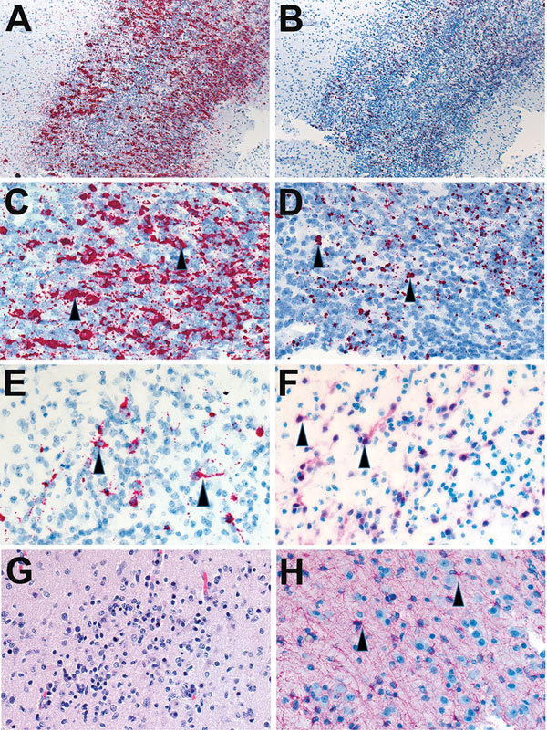
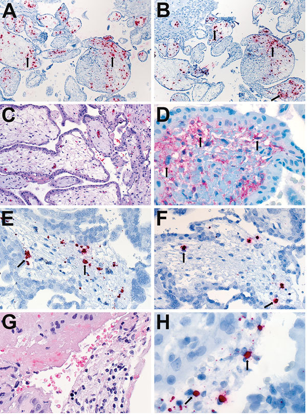
Zika virus is causally linked with congenital microcephaly and may be associated with pregnancy loss. However, the mechanisms of Zika virus intrauterine transmission and replication and its tropism and persistence in tissues are poorly understood. We tested tissues from 52 case-patients: 8 infants with microcephaly who died and 44 women suspected of being infected with Zika virus during pregnancy. By reverse transcription PCR, tissues from 32 (62%) case-patients (brains from 8 infants with microcephaly and placental/fetal tissues from 24 women) were positive for Zika virus. In situ hybridization localized replicative Zika virus RNA in brains of 7 infants and in placentas of 9 women who had pregnancy losses during the first or second trimester. These findings demonstrate that Zika virus replicates and persists in fetal brains and placentas, providing direct evidence of its association with microcephaly. Tissue-based reverse transcription PCR extends the time frame of Zika virus detection in congenital and pregnancy-associated infections.

Keywords: RT-PCR; Zika virus; brain; formalin-fixed; in-situ hybridization; paraffin-embedded tissues; placenta; replication; vector-borne infections; viruses.


Figures

Figure 1
Figure 1
Zika virus RNA load levels in human brain and placental tissues. The scatter plot graph shows the relative levels of Zika virus RNA in formalin-fixed, paraffin-embedded tissue sections, which were quantified by real-time quantitative reverse transcription PCR by using primer-probe sets for Zika virus envelope gene and β-actin mRNA. β-actin mRNA was used as an internal reference gene that provided a normalization factor for the amount of RNA extracted from a section. The copy number of Zika virus RNA per cell was calculated using β-actin mRNA copy number, which was estimated to be 1,500 copies/cell. The graph shows individual data points and superimposed horizontal lines at the geometric mean, and error bars show the 95% CI for that geometric mean. p values were calculated with nonparametric 1-way analysis of variance (Kruskal-Wallis test) followed by Dunn multiple comparison tests. The relative Zika virus RNA copy numbers for second/third trimester or full-term placentas were statistically significantly lower than those for first trimester placentas or infant brain tissues.
Figure 2
Figure 2
Localization of Zika virus RNA by in situ hybridization in brain tissues from infants with microcephaly. A) ISH with use of antisense probe. Zika virus genomic RNA (red stain) in cerebral cortex of an infant (case-patient no. 66, gestational age 26 wk). Original magnification ×10. B) ISH with use of sense probe. Serial section showing negative-strand replicative RNA intermediates (red stain) in the same areas shown in panel A. Original magnification ×10. C) ISH with use of antisense probe. Higher magnification of panel A, showing cytoplasmic staining of neural (arrowheads) and glial cells. Original magnification ×20. D) ISH with use of sense probe. Higher magnification of panel B, showing cytoplasmic staining of neural and glial cells (arrowheads). Original magnification ×20. E) ISH with use of antisense probe. Localization of negative-strand replicative RNA intermediates in neural cells or neurons (red, arrowheads) of another infant with fatal outcome (case-patient no. 67, gestational age 27 wk). Original magnification ×40. F) Immunostaining of neurons (arrowheads) with use of antibodies against neuronal nuclei in a serial section. Original magnification ×40. G) Hematoxylin and eosin stain showing cortical neural cells in a serial section. Original magnification ×40. H) Immunostaining of glial cells (arrowheads) with use of glial fibrillary acidic protein antibody in the same case. Original magnification ×40. ISH, in situ hybridization.
Figure 3
Figure 3
Localization of Zika virus RNA by ISH in placental tissues of women after spontaneous abortion. A) ISH with use of antisense probe. Zika virus genomic RNA localization in placental chorionic villi, predominantly within Hofbauer cells (red stain, arrows), of a case-patient who had spontaneous abortion at 11 wk gestation (case-patient no. 56). Original magnification ×10). B) ISH with use of sense probe. Serial section showing negative–strand replicative RNA intermediates (red stain, arrows) in the same cells shown in panel A. Original magnification ×10. C) Hematoxylin and eosin stain of placental tissue of a case-patient who experienced spontaneous abortion at 8 wk gestation (case-patient no. 47). Original magnification ×20. D) Immunostaining for CD163 highlighting villous Hofbauer cells in a serial section as seen in panel C. Original magnification ×63. E) ISH with use of antisense probe. Zika virus genomic RNA as seen in a serial section from the same case-patient as in panel C, showing staining within Hofbauer cells (red stain, arrows) of placental chorionic villi. Original magnification ×40. F) ISH with use of sense probe. Serial section showing negative-strand replicative RNA intermediates (red stain, arrows) in the same cells as shown in panel E. Original magnification ×40. G) Hematoxylin and eosin stain from the same case-patient as in panel C, showing inflammatory cell infiltrates in maternal side of placenta. Original magnification ×63. H) ISH with use of sense probe. Negative-strand replicative RNA intermediates (red stain, arrows) in inflammatory cells in a serial section. Original magnification ×63. ISH, in situ hybridization.

References

    1. Hennessey M, Fischer M, Staples JE. Zika virus spreads to new areas—region of the Americas, May 2015–January 2016. MMWR Morb Mortal Wkly Rep. 2016;65:55–8. 10.15585/mmwr.mm6503e1 - DOI - PubMed
    1. Rasmussen SA, Jamieson DJ, Honein MA, Petersen LR. Zika virus and birth defects—reviewing the evidence for causality. N Engl J Med. 2016;374:1981–7. 10.1056/NEJMsr1604338 - DOI - PubMed
    1. Cauchemez S, Besnard M, Bompard P, Dub T, Guillemette-Artur P, Eyrolle-Guignot D, et al. Association between Zika virus and microcephaly in French Polynesia, 2013-15: a retrospective study. Lancet. 2016;387:2125–32. 10.1016/S0140-6736(16)00651-6 - DOI - PMC - PubMed
    1. Simeone RM, Shapiro-Mendoza CK, Meaney-Delman D, Petersen EE, Galang RR, Oduyebo T, et al. ; Zika and Pregnancy Working Group. Possible Zika virus infection among pregnant women–United States and Territories, May 2016. MMWR Morb Mortal Wkly Rep. 2016;65:514–9. 10.15585/mmwr.mm6520e1 - DOI - PubMed
    1. Calvet G, Aguiar RS, Melo AS, Sampaio SA, de Filippis I, Fabri A, et al. Detection and sequencing of Zika virus from amniotic fluid of fetuses with microcephaly in Brazil: a case study. Lancet Infect Dis. 2016;16:653–60. 10.1016/S1473-3099(16)00095-5 - DOI - PubMed

LinkOut - more resources