. 2017 Apr;11(4):1029-1042.
doi: 10.1038/ismej.2016.163. Epub 2016 Dec 13.

Phage-inducible islands in the Gram-positive cocci

Affiliations

Phage-inducible islands in the Gram-positive cocci

Roser Martínez-Rubio et al. ISME J. 2017 Apr.

Abstract

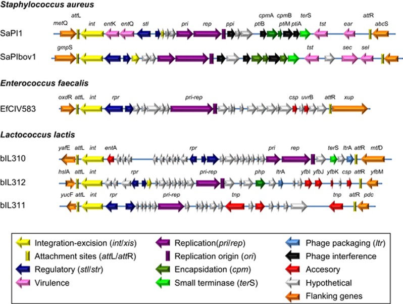
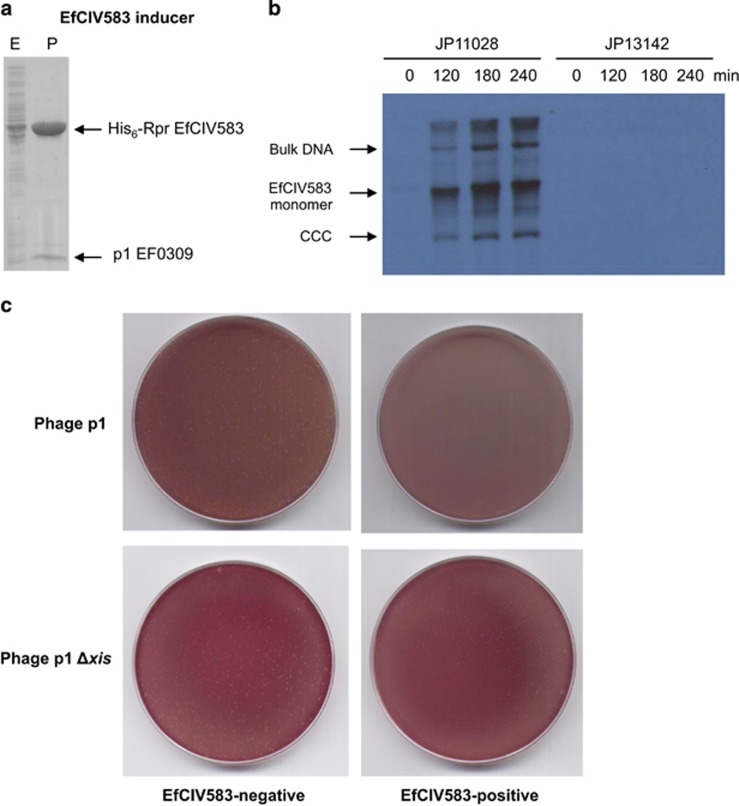
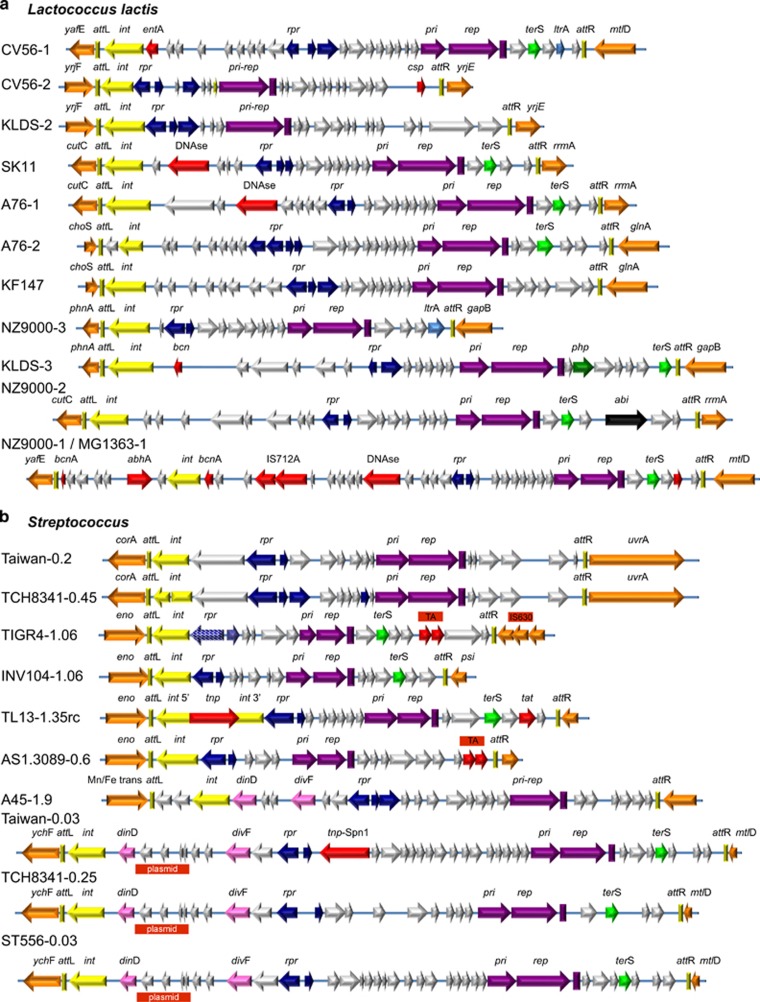
The SaPIs are a cohesive subfamily of extremely common phage-inducible chromosomal islands (PICIs) that reside quiescently at specific att sites in the staphylococcal chromosome and are induced by helper phages to excise and replicate. They are usually packaged in small capsids composed of phage virion proteins, giving rise to very high transfer frequencies, which they enhance by interfering with helper phage reproduction. As the SaPIs represent a highly successful biological strategy, with many natural Staphylococcus aureus strains containing two or more, we assumed that similar elements would be widespread in the Gram-positive cocci. On the basis of resemblance to the paradigmatic SaPI genome, we have readily identified large cohesive families of similar elements in the lactococci and pneumococci/streptococci plus a few such elements in Enterococcus faecalis. Based on extensive ortholog analyses, we found that the PICI elements in the four different genera all represent distinct but parallel lineages, suggesting that they represent convergent evolution towards a highly successful lifestyle. We have characterized in depth the enterococcal element, EfCIV583, and have shown that it very closely resembles the SaPIs in functionality as well as in genome organization, setting the stage for expansion of the study of elements of this type. In summary, our findings greatly broaden the PICI family to include elements from at least three genera of cocci.


Conflict of interest statement

The authors declare no conflict of interest.

Figures

Figure 1
Figure 1
Genome maps for PICIs and related elements. The originally identified PICIs from E. faecalis and L. lactis compared with SaPI1 and SaPIbov1. Additional details for these elements can be found in Supplementary Table S7.
Figure 2
Figure 2
Characterization of EfCIV583-encoded Int protein. (a) Schematic representation of the EfCIV583-dependent excision and circularization processes. The relevant genes, genetic markers and PCR primer binding sites are shown. (b) Detection of EfCIV583 excision and circularization. DNA from E. faecalis VE14089 was extracted and PCR-amplified using specific primers (see scheme in a) recognizing the external and internal sequence of the element (integration: I), primers recognizing the flanking sequences (excision: E) or PCR-amplified using a pair of primers set divergently at both termini of the island (circularization: C). M: molecular weight marker. (c) Constructs used for test site-specific integration in E. coli. Top: pCN51 derivatives containing the EfCIV583 attPI-int gene. Bottom: thermosensitive derivatives of pMAK700 carrying the attC from the E. faecalis chromosome. The relevant genetic markers and restriction enzyme sites are shown. (d) Plasmid DNA was isolated from overnight cultures grown at 37 °C (for strains carrying uniquely the pCN51 or pMAK700 derivatives) or at 43 °C (for strain carrying both plasmids), in the presence of ampicillin (pCN51) or chloramphenicol (pMAK700 derivative and cointegrative plasmid). Plasmids were digested with BamHI (pMAK700 derivatives) or with SalI (pCN51 derivatives and cointegrative plasmids).
Figure 3
Figure 3
Characterization of the EfCIV583-encoded Stl repressor. (a) Schematic representation of the different blaZ transcriptional fusions. The relevant genes are shown. (b) S. aureus RN4220 strains containing the plasmids represented in a were assayed for β-lactamase activity under standard conditions, or after MC induction. Samples were normalized for total cell mass. Values are presented are the averages (±s.d.) of three independent assays.
Figure 4
Figure 4
Induction of EfCIV583 by MC. MC (1 μg ml−1) was added to a culture of E. faecalis VE14089 (EfCIV583-positive) or E. faecalis VE14089 Δp1, followed by incubation at 32 °C. Samples were removed at the indicated time points and used to prepare minilysates, which were resolved on a 0.7% agarose gel (a), and Southern blotted with an EfCIV583 probe (b). In panel (c) is a stained gel and a Southern blot of DNA extracted from phage particles in a lysate of an MC-treated culture of VE14089. (d) The putative p1 pac site (colored in red) is embedded in the terS gene (colored in green). Its homolog in EfCIV583 is located between two genes (colored in blue). See text for explanation. Bottom: The predicted EfCIV583 and p1 pac sites are aligned using ClustalW2. A full-colour version of this figure is available at the ISME Journal Online.
Figure 5
Figure 5
Identification of the EfCIV583 inducer. (a) Affinity chromatography of p1 EF0309 using His6-RprEfCIV583. E. coli strain expressing the EF0309/His6-RprEfCIV583 pair was IPTG (isopropyl β-d-1-thiogalactopyranoside)-induced and, after disruption of the cells, the expressed proteins were applied to a Ni2+ agarose column and eluted. The presence of the different proteins was monitored in the load (lanes E), flow-through, wash and elute fractions (P) by Coomassie staining. The identity of the purified proteins was determined by in-gel enzymatic digestion and mass fingerprinting. It is assumed that the presence of Xis in the eluate represents an Rpr–Xis complex. (b) MC (1 μg ml−1) was added to a culture of E. faecalis JP11028 (EfCIV583/p1-positive) or E. faecalis JP13142 (JP11028 p1Δxis), followed by incubation at 32 °C. Samples were removed at the indicated time points and used to prepare minilysates, which were resolved on a 0.7% agarose gel, and Southern blotted with an EfCIV583 probe. (c) EfCIV583 interference with phage reproduction. Approximately 108 bacteria (carrying or not the EfCIV583 element) were infected with 400 plaque forming units (PFU) of phage φ1 or phage p1 Δxis, plated on phage bottom agar and incubated 24 h at 32 °C. Plates were stained with 0.1% triphenyl tetrazolium chloride in TSB (tryptic soy broth) media and photographed. CCC, closed circular form.
Figure 6
Figure 6
PICI genomes. (a) L. lactis PICI genomes identified by searching with b3IL10 genes, arranged by att sites. Also shown is a highly unusual element from L. lactis KF147, which may be related to integrative and conjugative element (ICE) elements and plasmids, as it has an integrase plus putative plasmid replication and segregation genes. It could be confused with a PICI, except that its transcriptional organization does not fit. (b) Genomes of PICIs of S. pneumoniae and other streptococci. The color coding of the PICI genes is the same as in Figure 1.

References

    1. Bolotin A, Wincker P, Mauger S, Jaillon O, Malarme K, Weissenbach J et al. (2001). The complete genome sequence of the lactic acid bacterium Lactococcus lactis ssp. lactis IL1403. Genome Res 11: 731–753. - PMC - PubMed
    1. Carpena N, Manning KA, Dokland T, Marina A, Penadés JR. (2016). Convergent evolution of pathogenicity islands in helper cos phage interference. Philos Trans R Soc Lond B 371: 20150505. - PMC - PubMed
    1. Charpentier E, Anton AI, Barry P, Alfonso B, Fang Y, Novick RP. (2004). Novel cassette-based shuttle vector system for gram-positive bacteria. Appl Environ Microbiol 70: 6076–6085. - PMC - PubMed
    1. Chen J, Novick RP. (2009). Phage-mediated intergeneric transfer of toxin genes. Science 323: 139–141. - PubMed
    1. Chen J, Carpena N, Quiles-Puchalt N, Ram G, Novick RP, Penadés JR. (2015. a). Intra- and inter-generic transfer of pathogenicity island-encoded virulence genes by cos phages. ISME J 9: 1260–1263. - PMC - PubMed

MeSH terms