. 2017 Jan;50(1):310-316.
doi: 10.3892/ijo.2016.3798. Epub 2016 Dec 9.

Microtubule actin cross-linking factor 1, a novel target in glioblastoma

Affiliations

Microtubule actin cross-linking factor 1, a novel target in glioblastoma

Najlaa Afghani et al. Int J Oncol. 2017 Jan.

Abstract

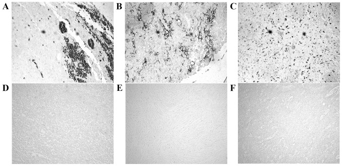
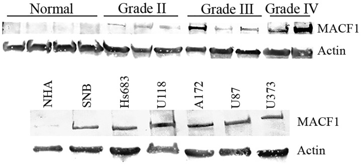
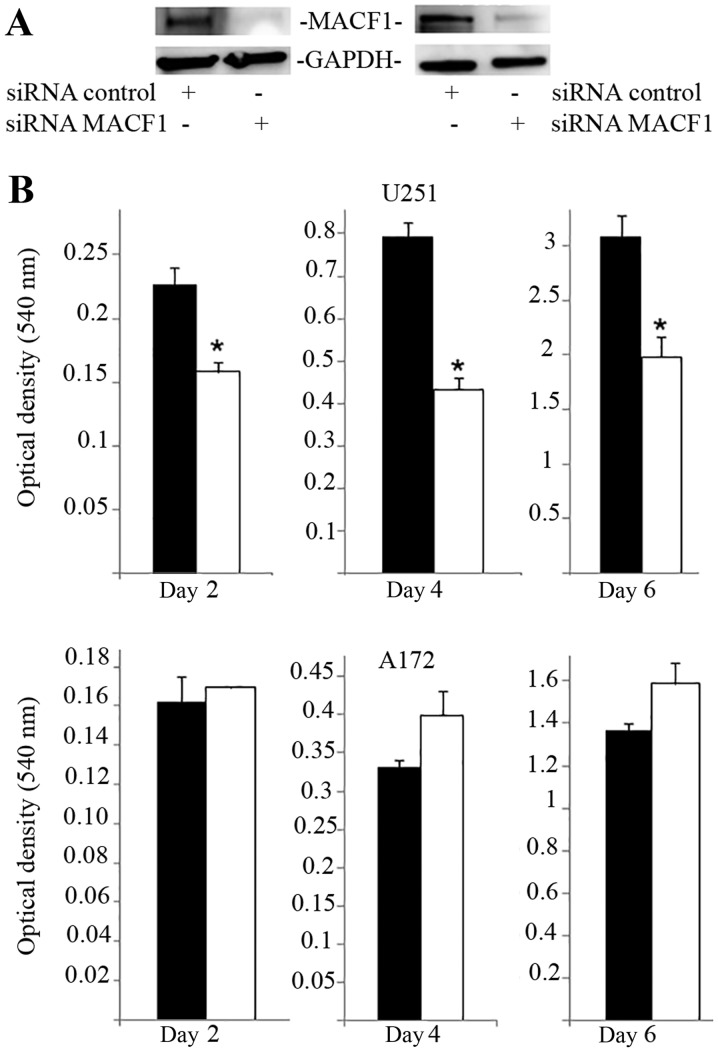
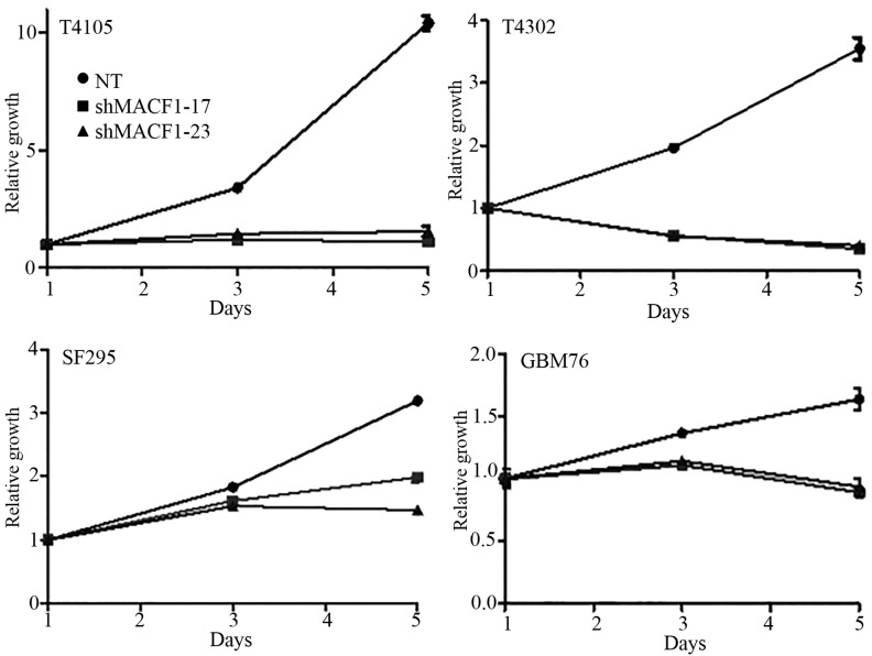
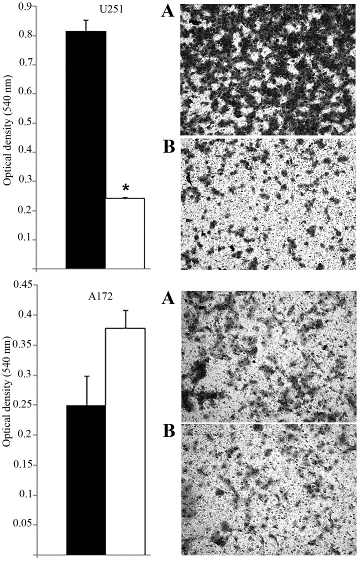
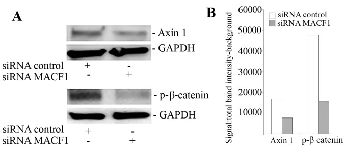
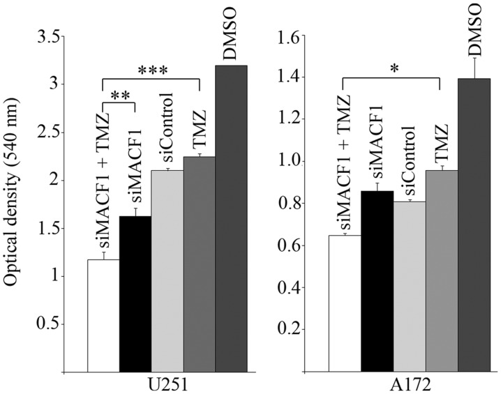
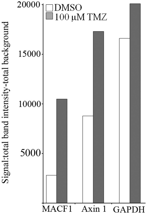
Genetic heterogeneity is recognized as a major contributing factor of glioblastoma resistance to clinical treatment modalities and consequently low overall survival rates. This genetic diversity results in variations in protein expression, both intratumorally and between individual glioblastoma patients. In this regard, the spectraplakin protein, microtubule actin cross-linking factor 1 (MACF1), was examined in glioblastoma. An expression analysis of MACF1 in various types of brain tumor tissue revealed that MACF1 was predominately present in grade III-IV astroctyomas and grade IV glioblastoma, but not in normal brain tissue, normal human astrocytes and lower grade brain tumors. Subsequent genetic inhibition experiments showed that suppression of MACF1 selectively inhibited glioblastoma cell proliferation and migration in cell lines established from patient derived xenograft mouse models and immortalized glioblastoma cell lines that were associated with downregulation of the Wnt-signaling mediators, Axin1 and β-catenin. Additionally, concomitant MACF1 silencing with the chemotherapeutic agent temozolomide (TMZ) used for the clinical treatment of glioblastomas cooperatively reduced the proliferative capacity of glioblastoma cells. In conclusion, the present study represents the first investigation on the functional role of MACF1 in tumor cell biology, as well as demonstrates its potential as a unique biomarker that can be targeted synergistically with TMZ as part of a combinatorial therapeutic approach for the treatment of genetically multifarious glioblastomas.


Figures

Figure 1
Figure 1
MACF1 expression in brain tumors and normal brain tissue. MACF1 was robustly expressed in glioblastoma (A–C) but not detected in oligodendroglioma (D), medulloblastoma (E), or normal brain tissue (F). MACF1 protein is labeled as black diaminobenzidine (DAB) positive cells. Displayed is an immunohistochemistry labeling experiment representative of three independent experiments that showed similar results (total magnification, ×200).
Figure 2
Figure 2
Western blot analysis of MACF1 expression in astrocytomas and glioblastoma cell lines. Astrocytoma tissue taken from tumor patients (top panel) and glioblastoma cell lines (bottom panel) displayed increased MACF1 protein levels as compared to normal human astrocytes (NHA) and normal brain tissue. Immunoblots are representative of at least four experiments that showed equivalent results. Actin was used as a loading control to assess that lanes were loaded with the same amount of total proteins.
Figure 3
Figure 3
Proliferative response of glioblastoma cells to MACF1 inhibition. (A) Western blot analysis of U251 and A172 cells treated for six days with MACF1 siRNAs nearly abolished MACF1 expression in U251 and A172 cells. (B) Bar graphs show that reduction in MACF1 levels was accompanied with a significant decrease in the proliferative capacity of U251 cells (*P<0.05 as determined by the Student's t-test; error bars, SE) but not A172 cells. Data shown are representative of at least three independent experiments performed in duplicate. Black bars, control siRNA treated cells; white bars, MACF1 siRNA treated cells.
Figure 4
Figure 4
Downregulation of MACF1 impairs proliferation of patient derived xenograft cell lines. Time-course analysis showed that inhibition of MACF1 with shRNAs decreased the proliferative capacity of patient derived glioblastoma cell lines established in xenograft mouse models.
Figure 5
Figure 5
Effects of silencing MACF1 on glioblastoma cell migration. Downregulation of MACF1 significantly impeded cell migration of U251 cells (top panel), but not A172 cells (bottom panel). Bar graphs displayed are representative of three independent experiments performed in duplicate that showed similar results. Black bars, control siRNA treated cells; white bars, MACF1 siRNA treated cells (*P<0.05 as determined by the Student's t-test; error bars, SE).
Figure 6
Figure 6
Suppression of MACF1 reduced Axin1 and β-catenin expression. Inhibition of MACF1 with siRNAs downregulated Axin1 and β-catenin protein levels in U251 glioblastoma cells. (A) Western blot experiments and (B) densitometric analysis. Data shown are representative of at least three independent experiments that displayed comparative results.
Figure 7
Figure 7
MACF1 repression with concurrent TMZ treatment. Glioblastoma cells with silenced MACF1 treated with 100 µM TMZ led to a decrease in glioblastoma cell proliferation compared to the treatment with vehicle DMSO, control siRNAs, TMZ, or MACF1 siRNA alone as determined by ANOVA analysis and Sidak's post hoc multiple comparisons test (*P<0.05, **P<0.01, ***P<0.001).
Figure 8
Figure 8
Induction of MACF1 and Axin1 by TMZ. Densitometric analysis of immunoblot showed that TMZ upregulated MACF1 and Axin1 protein levels in U251 glioblastoma cells. Data shown are representative of at least three independent experiments that displayed comparative results.

References

    1. Ostrom QT, Bauchet L, Davis FG, Deltour I, Fisher JL, Langer CE, Pekmezci M, Schwartzbaum JA, Turner MC, Walsh KM, et al. The epidemiology of glioma in adults: A 'state of the science' review. Neuro Oncol. 2014;16:896–913. doi: 10.1093/neuonc/nou087. - DOI - PMC - PubMed
    1. Reardon DA, Wen PY. Glioma in 2014: Unravelling tumour heterogeneity - implications for therapy. Nat Rev Clin Oncol. 2015;12:69–70. doi: 10.1038/nrclinonc.2014.223. - DOI - PubMed
    1. Parsons DW, Jones S, Zhang X, Lin JC, Leary RJ, Angenendt P, Mankoo P, Carter H, Siu IM, Gallia GL, et al. An integrated genomic analysis of human glioblastoma multiforme. Science. 2008;321:1807–1812. doi: 10.1126/science.1164382. - DOI - PMC - PubMed
    1. Verhaak RG, Hoadley KA, Purdom E, Wang V, Qi Y, Wilkerson MD, Miller CR, Ding L, Golub T, Mesirov JP, et al. Cancer Genome Atlas Research Network Integrated genomic analysis identifies clinically relevant subtypes of glioblastoma characterized by abnormalities in PDGFRA, IDH1, EGFR, and NF1. Cancer Cell. 2010;17:98–110. doi: 10.1016/j.ccr.2009.12.020. - DOI - PMC - PubMed
    1. Heimberger AB, Hlatky R, Suki D, Yang D, Weinberg J, Gilbert M, Sawaya R, Aldape K. Prognostic effect of epidermal growth factor receptor and EGFRvIII in glioblastoma multiforme patients. Clin Cancer Res. 2005;11:1462–1466. doi: 10.1158/1078-0432.CCR-04-1737. - DOI - PubMed