Review
. 2017 Jan 6:57:19-37.
doi: 10.1146/annurev-pharmtox-010716-104710. Epub 2016 Dec 7.

Nanobodies to Study G Protein-Coupled Receptor Structure and Function

Affiliations
Review

Nanobodies to Study G Protein-Coupled Receptor Structure and Function

Aashish Manglik et al. Annu Rev Pharmacol Toxicol. .

Abstract

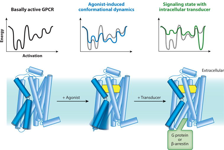
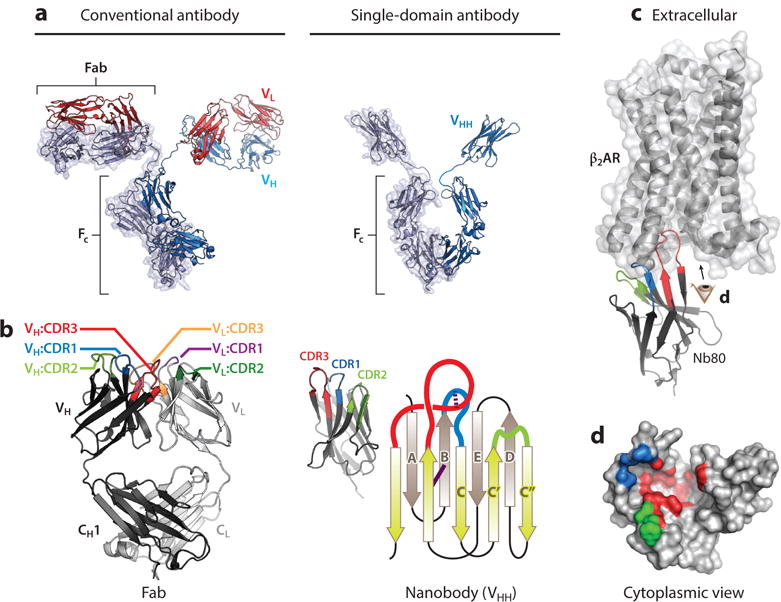
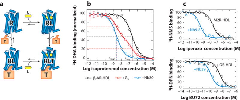
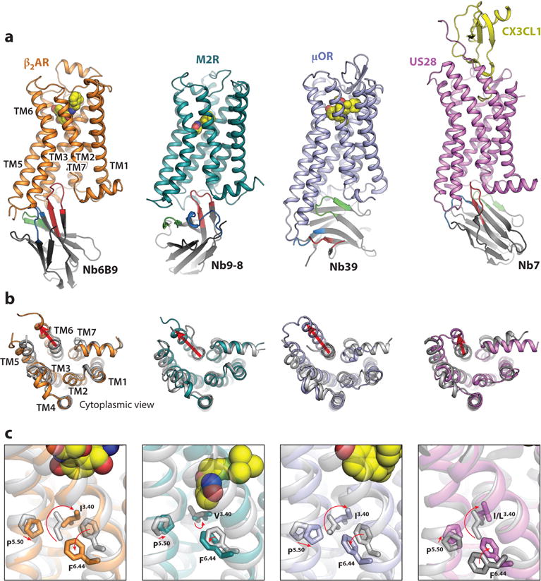
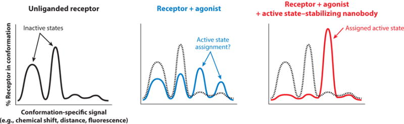
Ligand-induced activation of G protein-coupled receptors (GPCRs) is a key mechanism permitting communication between cells and organs. Enormous progress has recently elucidated the structural and dynamic features of GPCR transmembrane signaling. Nanobodies, the recombinant antigen-binding fragments of camelid heavy-chain-only antibodies, have emerged as important research tools to lock GPCRs in particular conformational states. Active-state stabilizing nanobodies have elucidated several agonist-bound structures of hormone-activated GPCRs and have provided insight into the dynamic character of receptors. Nanobodies have also been used to stabilize transient GPCR transmembrane signaling complexes, yielding the first structural insights into GPCR signal transduction across the cellular membrane. Beyond their in vitro uses, nanobodies have served as conformational biosensors in living systems and have provided novel ways to modulate GPCR function. Here, we highlight several examples of how nanobodies have enabled the study of GPCR function and give insights into potential future uses of these important tools.

Keywords: G protein–coupled receptor; conformational plasticity; crystallographic chaperone; intrabody; nanobody; receptor activation.


Figures

Figure 1
Figure 1
Conformational complexity in G protein-coupled receptor (GPCR) function. Unliganded and antagonist-bound GPCRs are conformationally dynamic. Agonists further induce receptor dynamics to varying degrees, and the crystallographically observed active state is stabilized only in the presence of a G protein or an active state-stabilizing chaperone (49). Energy diagrams illustrate the conformational complexity of GPCRs and highlight the inherent difficulty in capturing agonist-bound receptors in conformationally homogeneous crystallographic lattices.
Figure 2
Figure 2
Nanobody structure and function and comparison to conventional antibodies. (a) Comparison of conventional antibodies to camelid single-domain antibodies. Conventional antibodies are heterotetrameric molecules consisting of two heavy chains (VH) and two light chains (VL) with a conserved domain called the crystallizable fragment (FC). Variable loops responsible for antigen binding are within the distal tips of the Fab domain. Camelid single-chain antibodies contain a single immunoglobulin domain (VHH) that binds antigens individually. (b) Comparison of the minimal binding domain of conventional antibodies (Fab) and single-domain antibodies (VHH or nanobody). The antigen-binding region of a Fab is composed of six complementarity-determining regions (CDRs), with three in each VH and VL. Correct VH/VL pairing is required for antigen binding. In contrast, nanobodies contain three CDRs, and the single immunoglobulin fold is sufficient for antigen binding. The nanobody immunoglobulin fold is built from a pair of antiparallel β sheets with a conserved disulfide bond (solid purple line). The CDRs originate from loops between individual strands. Many nanobodies contain an extra interloop disulfide bond that restricts the flexibility of CDR1 and CDR3 (dotted purple line). (c) The prolate structure of the nanobody forms a convex paratope surface, which allows it to access antigenic cavities. In the β2-adrenergic receptor·Nanobody80 (β2AR·Nb80) complex shown here [Protein Data Bank (PDB) ID: 3P0G], CDR3 of Nb80 inserts into the cytoplasmic surface of active β2AR, with additional interactions made by CDR1 and CDR2. The resulting β2AR epitope recognized by Nb80, viewed from the cytoplasmic surface (eye symbol), is displayed in panel d. Note that each CDR binds different regions of the complex three-dimensional epitope that is discontinuous in β2AR sequence. CDRs and framework residues in this figure have been defined according to the International ImMunoGeneTics Information System (IMGT) (138).
Figure 3
Figure 3
Active state-stabilizing nanobodies recapitulate the G protein-coupled receptor ternary complex model. (a) In the ternary complex model, agonist (L) binding results in increased affinity for the intracellular transducer, most commonly one of the heterotrimeric G proteins. Conversely, binding of a transducer (T) to the receptor (R) induces a reciprocal increase in agonist affinity. (b) The β2-adrenergic receptor (β2AR) reconstituted into high-density lipoprotein (HDL) particles shows a monophasic competition curve resulting from its affinity for the agonist isoproterenol. Addition of Gs results in a biphasic curve, with a fraction of receptor displaying an increase in agonist affinity induced by the G protein. Nanobody80 (Nb80), an active state-stabilizing nanobody for the β2AR, induces a similar high-affinity agonist state. (c) Similar nanobodies were identified subsequently for the M2 muscarinic receptor (M2R) (Nb9-8) and the μ-opioid receptor (μOR) (Nb39). In each case, active state-stabilizing nanobodies increase the affinity of agonists. Other abbreviations: 3H-DHA, 3H-dihydroalprenolol; 3H-DPN, 3H-diprenorphine; 3H-NMS, 3H-N-methylscopolamine.
Figure 4
Figure 4
Structural basis of G protein-coupled receptor (GPCR) activation revealed by nanobody-assisted crystallography. (a) Active-state structures of agonist-bound, nanobody-stabilized GPCRs. Each nanobody binds the intracellular surface of the receptor in a unique orientation. Whereas complementarity-determining region 3 (CDR3) (red) most commonly extends into the core of the receptor, Nanobody39 (Nb39) uses framework residues, CDR1, and CDR2 to engage with the μ-opioid receptor (μOR). (b) Cytoplasmic view comparing inactive [grey; Protein Data Bank (PDB) IDs: 2RH1, 3UON, 4DKL, 4XT1] and active receptors (colored; PDB IDs: 4LDE, 4MQS, 5C1M, 4MBS). Key conserved features include an outward displacement of transmembrane helix 6 (TM6), an inward movement of TM5, and a rearrangement of TM7. (c) Active-state GPCR structures reveal a conserved mode of allostery between the ligand-binding pocket and the cytoplasmic domain. The agonists (yellow spheres) induce conformational changes in the ligand-binding pocket, which are relayed via a conserved set of transmission switch residues (F6.44, P5.50, and I/L/V3.40) that undergo similar rearrangements upon activation.
Figure 5
Figure 5
Nanobody-based assignment of G protein-coupled receptor conformational states. In many spectroscopic experiments, distinct signals arise from unique conformations of a given receptor. In the absence of ligand, multiple signals reflect conformational heterogeneity. Addition of agonists is associated with increased conformational heterogeneity and the presence of new signals arising from a greater number of conformations. Assignment of a specific signal to a crystallographically observed conformation or a pharmacologically characterized state is challenging (middle panel). Active state-stabilizing nanobodies can conformationally stabilize the receptor, thereby allowing clear assignment of signals and further interpretation of spectroscopic data.

Similar articles

Cited by

References

    1. Lagerström MC, Schioth HB. Structural diversity of G protein-coupled receptors and significance for drug discovery. Nat Rev Drug Discov. 2008;7:339–57. - PubMed
    1. Lefkowitz RJ. Historical review: a brief history and personal retrospective of seven-transmembrane receptors. Trends Pharmacol Sci. 2004;25:413–22. - PubMed
    1. Palczewski K, Kumasaka T, Hori T, Behnke CA, Motoshima H, et al. Crystal structure of rhodopsin: a G protein-coupled receptor. Science. 2000;289:739–45. - PubMed
    1. Katritch V, Cherezov V, Stevens RC. Structure-function of the G protein-coupled receptor superfamily. Annu Rev Pharmacol Toxicol. 2013;53:531–56. - PMC - PubMed
    1. Pierce KL, Premont RT, Lefkowitz RJ. Seven-transmembrane receptors. Nat Rev Mol Cell Biol. 2002;3:639–50. - PubMed

Publication types

MeSH terms