Review
. 2017 Jan 24:12:25-52.
doi: 10.1146/annurev-pathol-012615-044145. Epub 2016 Dec 5.

Complement Dysregulation and Disease: Insights from Contemporary Genetics

Affiliations
Review

Complement Dysregulation and Disease: Insights from Contemporary Genetics

M Kathryn Liszewski et al. Annu Rev Pathol. .

Abstract

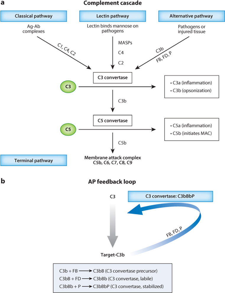
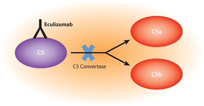
The vertebrate complement system consists of sequentially interacting proteins that provide for a rapid and powerful host defense. Nearly 60 proteins comprise three activation pathways (classical, alternative, and lectin) and a terminal cytolytic pathway common to all. Attesting to its potency, nearly half of the system's components are engaged in its regulation. An emerging theme over the past decade is that variations in these inhibitors predispose to two scourges of modern humans. One, occurring most often in childhood, is a rare but deadly thrombomicroangiopathy called atypical hemolytic uremic syndrome. The other, age-related macular degeneration, is the most common form of blindness in the elderly. Their seemingly unrelated clinical presentations and pathologies share the common theme of overactivity of the complement system's alternative pathway. This review summarizes insights gained from contemporary genetics for understanding how dysregulation of this powerful innate immune system leads to these human diseases.

Keywords: C3; C3 glomerulopathies; CD46; age-related macular degeneration; alternative complement pathway; atypical hemolytic uremic syndrome; factor B; factor H; factor I.


Figures

Figure 1
Figure 1
The complement cascades. (a) The three pathways of complement activation are shown. Although each is independently triggered, they all merge at the step of C3 activation. The classical pathway (CP) is initiated by the binding of antibody (Ab) to antigen (Ag) and the lectin pathway (LP) by the binding of lectin to an oligosaccharide. The alternative pathway (AP) turns over continuously, generating small amounts of 3(H2O) (the thioester-hydrolyzed form of C3) and C3b, which it amplifies in the presence of pathogens or injured tissue. C3(H2O) behaves like C3b in that it can bind factor B (FB) to initiate the AP (see below). Activation of the complement system leads to inflammation (release of anaphylatoxins C3a and C5a), opsonization (the coating of targets with C3b and/or C4b), and membrane perturbation (formation of the membrane attack complex, MAC). (b) The AP’s feedback loop of complement activation. First, C3b attaches to the surface of a target, such as a pathogen or self-debris. Next, C3b binds to FB, and then factor D (FD) cleaves FB to form the AP C3 convertase (i.e., an enzyme that cleaves C3 to C3b). This convertase is stabilized by the binding of properdin (P). The C3 convertase triggers the amplification loop via the generation of more C3b. The initial C3b can be generated by the CP and LP or tick over [formation of C3(H2O), which can also bind FB to trigger the AP]. Abbreviations: MASPs, mannose-binding lectin-associated serine proteases; C3(H2O), C3 with a cleaved thioester bond. Modified with permission from Liszewski MK, Atkinson JP. Complement Pathways. In UpToDate, Post TW (Ed), UpToDate, Waltham, MA, accessed on 4–10-15. Copyright © 2016 UpToDate, Inc.
Figure 2
Figure 2
Complement regulation: cofactor activity. In Step 1 of this example, C3b becomes covalently bound to a target (e.g., an endothelial cell). In Step 2, the membrane-anchored host protein, CD46, locates and binds to the C3b. In Step 3, the plasma serine protease factor I (FI) binds to the C3b/cofactor protein complex. In Step 4, FI proteolytically cleaves C3b to iC3b (a product that cannot participate in the feedback loop) and C3f (the small degradation fragment) and thereby halts further complement activation. Modified with permission from Liszewski & Atkinson (22). Published by BioMed Central © 2015.
Figure 3
Figure 3
Cross-sectional diagrams of the human eye. (a) Macroscopic schematic picture of the human eye. (b) Microscopic schematic picture of the retina in health and in age-related macular degeneration (AMD). Subretinal drusen accumulate; this accumulation may block nutrients and thereby damage the photoreceptor cell layer, leading to atrophy. In theory, the primary process in AMD could be excessive photoreceptor damage, retinal pigment epithelial dysfunction, an alteration in Bruch’s membrane, or vascular damage to the choroid. Modified from Schramm et al. (34). Published by Elsevier © 2014.
Figure 4
Figure 4
Factor H (FH) in age-related macular degeneration (AMD) and atypical hemolytic uremic syndrome (aHUS). FH is a 155-kDa plasma protein composed of 20 repeating units, called complement control proteins (CCPs), consisting of approximately 60 amino acids, linked together by 3–8 amino acids. The sites involved in ligand binding and attachment to membranes bearing glycosaminoglycans (GAGs) are identified. A common variant (Y402H) associated with AMD risk is located in the seventh CCP. The C terminus of FH (repeats 19 and 20) is particularly associated with aHUS, as 60% of all FH mutants occur in these two CCPs, which mediate both ligand and GAG binding. In contrast, mutations causing a regulatory dysfunction in CCPs 1–4 are commonly observed in both aHUS and AMD. Abbreviations: CA, cofactor activity; DAA, decay-accelerating activity; SNP, single-nucleotide polymorphism. Modified from Schramm et al. (34). Published by Elsevier © 2014.
Figure 5
Figure 5
Disease-associated membrane cofactor protein (MCP) (CD46) mutations. The schematic depicts the CD46 protein, genomic organization, and disease-associated mutations. CD46 has a 34-amino acid signal peptide (SP). The mature protein consists of four complement control protein (CCP) repeats that house the sites for its regulatory cofactor activity. This is followed by an alternatively spliced region for O-glycosylation (segments A, B, C), a short 12-amino acid segment of undefined function (U), a transmembrane domain (TM), and one of two alternatively spliced cytoplasmic tails (CYT-1 or CYT-2). The gene consists of 14 exons and 13 introns for a minimum length of 43 kb. More than 90% of the mutations are rare (gene frequency less than 1%) and deleterious. Note that in a majority, the protein is not synthesized (splice site, stop, and cysteine mutations) or secreted, whereas in the others, it is expressed but dysfunctional (commonly missense mutations). A majority of mutations for aHUS and for other disorders (such as systemic sclerosis, systemic lupus erythematosus, and pregnancy-related disorders) occur in the four CCPs. Mutations and their locations are listed below the exons. Modified with permission from Liszewski & Atkinson (22). Published by BioMed Central © 2015.
Figure 6
Figure 6
Schematic diagram of the protein structure of factor I (FI). FI is synthesized as a single polypeptide chain of 90 kDa. Following removal of the signal peptide (SP), FI is proteolytically processed into a heavy and a light chain linked by a disulfide bond. The noncatalytic heavy chain consists of several modules that share homology to the FI membrane attack complex domain (FIMAC), the scavenger receptor cysteine-rich domain (SRCR), and two low-density lipoprotein receptor domains (LDLR1 and LDLR2). The light chain comprises the serine protease domain that contains the expected catalytic triad of His-362, Asp-411, and Ser-507, responsible for its cleavage activities. Modified from Schramm et al. (34). Published by Elsevier © 2014
Figure 7
Figure 7
Protein structure of C3. (a) C3 is synthesized as a single precursor chain. Following proteolytic processing, the mature structure of C3 consists of two disulfide-linked polypeptide chains (α and β). Features include eight macroglobulin domains (MG) with approximately 100 amino acids in each and a linker domain (LNK). The N terminus of the α-chain consists of an anaphylatoxin domain (ANA), an αNT module, a CUB domain (C1r/C1s, Uefg, Bmp1), a thioester-containing domain (TED) allowing for C3 attachment to a target, an anchor domain, and a C-terminal C345C segment. Modified with permission from Schramm et al. (34) and published by Elsevier © 2014. (b) Surface representation of the three-dimensional structures of C3b bound by membrane cofactor protein (MCP) (blue) and factor H (FH) (orange). Abbreviation: CCP, complement control protein. Reprinted from Forneris et al. (30) and published by Wiley © 2016.
Figure 8
Figure 8
Schematic diagram of the protein structure of factor B. Factor B is a mosaic glycoprotein composed of three types of protein modules. The N-terminal region (the Ba fragment) features three complement control protein (CCP) modules. This is followed by a von Willebrand factor type A (VWA) domain (a ligand and metal-binding site). The C terminus is a serine protease domain similar to that of trypsin. The capacity to cleave C3 is acquired through C3 convertase assembly (C3bBb) (See Figure 1b).
Figure 9
Figure 9
Therapeutic inhibition of C5 by eculizumab. Eculizumab is a humanized recombinant murine monoclonal antibody approved for treatment of paroxysmal nocturnal hemoglobinuria and atypical hemolytic uremic syndrome. The antibody binds C5 near the cleavage site and thereby prevents its enzymatic activation by the C5 convertase into C5b and the proinflammatory peptide C5a. The upstream immune-enhancing and autoimmune protective functions of C3 remain intact with this agent.

Similar articles

Cited by

References

    1. Gros P, Milder FJ, Janssen BJ. 2008. Complement driven by conformational changes. Nat. Rev. Immunol 8:48–58 - PubMed
    1. Zhu Y, Thangamani S, Ho B, Ding JL. 2005. The ancient origin of the complement system. EMBO J 24:382–94 - PMC - PubMed
    1. Nonaka M, Kimura A. 2006. Genomic view of the evolution of the complement system. Immunogenetics 58:701–13 - PMC - PubMed
    1. Walport MJ. 2001. Complement. First of two parts. N. Engl. J. Med 344:1058–66 - PubMed
    1. Chaplin H, Nasongkla M, Monroe MC. 1981. Quantitation of red blood cell–bound C3d in normal subjects and random hospitalized patients. Br. J. Haematol 48(1):69–78 - PubMed

MeSH terms

Substances