Review
. 2017 Jan 24:12:103-130.
doi: 10.1146/annurev-pathol-052016-100247. Epub 2016 Dec 5.

Necroptosis: Mechanisms and Relevance to Disease

Affiliations
Review

Necroptosis: Mechanisms and Relevance to Disease

Lorenzo Galluzzi et al. Annu Rev Pathol. .

Abstract

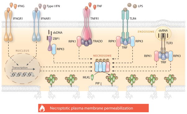
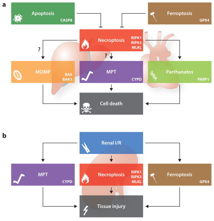
Necroptosis is a form of regulated cell death that critically depends on receptor-interacting serine-threonine kinase 3 (RIPK3) and mixed lineage kinase domain-like (MLKL) and generally manifests with morphological features of necrosis. The molecular mechanisms that underlie distinct instances of necroptosis have just begun to emerge. Nonetheless, it has already been shown that necroptosis contributes to cellular demise in various pathophysiological conditions, including viral infection, acute kidney injury, and cardiac ischemia/reperfusion. Moreover, human tumors appear to obtain an advantage from the downregulation of key components of the molecular machinery for necroptosis. Although such an advantage may stem from an increased resistance to adverse microenvironmental conditions, accumulating evidence indicates that necroptosis-deficient cancer cells are poorly immunogenic and hence escape natural and therapy-elicited immunosurveillance. Here, we discuss the molecular mechanisms and relevance to disease of necroptosis.

Keywords: caspases; damage-associated molecular patterns; immunogenic cell death; inflammation; mitochondrial permeability transition; necrostatin-1.


Figures

Figure 1
Figure 1
Molecular mechanisms of necroptosis. Necroptosis critically depends on the receptor-interacting serine-threonine protein kinase 3 (RIPK3)–mediated phosphorylation of mixed lineage kinase domain-like (MLKL), resulting in MLKL oligomerization, translocation to the inner leaflet of the plasma membrane, and cell death. The formation of the RIPK3- and MLKL-containing complex that precipitates necroptosis, the so-called necrosome, can be elicited by extracellular signals (such as the ligation of death receptors) as well as by intracellular cues (such as the presence of viral nucleic acids) and is regulated by a complex network of physical and functional protein-to-protein interactions. The best characterized signal transduction cascade resulting in necroptotic cell death is initiated by TNFR1 (official name: TNFRSF1A, tumor necrosis factor receptor superfamily member 1A) ligation in the presence of caspase inhibitors and/or SMAC (official name: DIABLO, diablo IAP-binding mitochondrial protein) mimetics and critically depends on the phosphorylation of RIPK3 by RIPK1. In several other circumstances, however, RIPK1 is dispensable for necroptotic responses or even inhibits them in an active manner. This applies to various other TNFR1 interactors that participate in necroptotic signaling, most of which also regulate caspase 8 (CASP8)-dependent apoptosis and proinflammatory NF-κB activation. Please note that several physical or functional interactions have been omitted for the sake of simplicity. Abbreviations: dsDNA, double-stranded DNA; dsRNA, double-stranded RNA; IFN, interferon; IFNAR1, interferon (alpha and beta) receptor 1; IFNG, interferon gamma; IFNGR1, interferon gamma receptor 1; LPS, lipopolysaccharide; P, phosphate; PIP, phosphatidylinositol phosphate; TLR, Toll-like receptor; TNF, tumor necrosis factor; TRADD, TNFRSF1A associated via death domain; TRIF (official name TICAM1), toll-like receptor adaptor molecule 1; ZBP1, Z-DNA binding protein 1.
Figure 2
Figure 2
Cross talk between necroptosis and other forms of regulated cell death. The signal transduction cascades that precipitate necroptosis and other forms of regulated cell death are highly intertwined. A heterotrimeric complex containing caspase 8 (CASP8), Fas associated via death domain (FADD), and the long isoform of cFLIP (official name CFLAR, CASP8 and FADD like apoptosis regulator), in which CASP8 retains partial proteolytic activity, robustly inhibits necroptosis, most likely owing to the CASP8-dependent cleavage of receptor-interacting serine-threonine kinase 1 (RIPK1), RIPK3, and cylindromatosis (CYLD). According to some reports that await confirmation, RIPK1 and/or RIPK3 may also mediate lethal effects by precipitating mitochondrial outer membrane permeabilization (MOMP), mitochondrial permeability transition (MPT)-driven regulated necrosis, or parthanatos. Along similar lines, the endogenous inhibitor of ferroptosis glutathione peroxidase 4 (GPX4) may suppress necroptosis by preserving optimal CASP8 functions (a). In specific situations, however, it seems that the signal transduction cascades precipitating necroptosis, MPT-driven regulated necrosis, ferroptosis, and parthanatos operate in a manner that is completely independent from each other. One of these scenarios is represented by renal ischemia/reperfusion (I/R) (b). Abbreviations: BAK1, BCL2-antagonist/killer 1; BAX, BCL2-associated X protein; CYPD (official name, PPIF), peptidylprolyl isomerase F; MLKL, mixed lineage kinase domain-like; PARP1, poly(ADP-ribose) polymerase 1.

References

    1. Galluzzi L, Bravo-San Pedro JM, Vitale I, Aaronson SA, Abrams JM, et al. Essential versus accessory aspects of cell death: recommendations of the NCCD 2015. Cell Death Differ. 2015;22:58–73. - PMC - PubMed
    1. Galluzzi L, Bravo-San Pedro JM, Kroemer G. Organelle-specific initiation of cell death. Nat Cell Biol. 2014;16:728–36. - PubMed
    1. Green DR, Galluzzi L, Kroemer G. Cell biology. Metabolic control of cell death Science. 2014;345:1250256. - PMC - PubMed
    1. Fuchs Y, Steller H. Programmed cell death in animal development and disease. Cell. 2011;147:742–58. - PMC - PubMed
    1. Sica V, Galluzzi L, Bravo-San Pedro JM, Izzo V, Maiuri MC, Kroemer G. Organelle-specific initiation of autophagy. Mol Cell. 2015;59:522–39. - PubMed

Substances