. 2016 Dec 13;7(6):e01721-16.
doi: 10.1128/mBio.01721-16.

The Conserved Coronavirus Macrodomain Promotes Virulence and Suppresses the Innate Immune Response during Severe Acute Respiratory Syndrome Coronavirus Infection

Affiliations

The Conserved Coronavirus Macrodomain Promotes Virulence and Suppresses the Innate Immune Response during Severe Acute Respiratory Syndrome Coronavirus Infection

Anthony R Fehr et al. mBio. .

Abstract

ADP-ribosylation is a common posttranslational modification that may have antiviral properties and impact innate immunity. To regulate this activity, macrodomain proteins enzymatically remove covalently attached ADP-ribose from protein targets. All members of the Coronavirinae, a subfamily of positive-sense RNA viruses, contain a highly conserved macrodomain within nonstructural protein 3 (nsp3). However, its function or targets during infection remain unknown. We identified several macrodomain mutations that greatly reduced nsp3's de-ADP-ribosylation activity in vitro Next, we created recombinant severe acute respiratory syndrome coronavirus (SARS-CoV) strains with these mutations. These mutations led to virus attenuation and a modest reduction of viral loads in infected mice, despite normal replication in cell culture. Further, macrodomain mutant virus elicited an early, enhanced interferon (IFN), interferon-stimulated gene (ISG), and proinflammatory cytokine response in mice and in a human bronchial epithelial cell line. Using a coinfection assay, we found that inclusion of mutant virus in the inoculum protected mice from an otherwise lethal SARS-CoV infection without reducing virus loads, indicating that the changes in innate immune response were physiologically significant. In conclusion, we have established a novel function for the SARS-CoV macrodomain that implicates ADP-ribose in the regulation of the innate immune response and helps to demonstrate why this domain is conserved in CoVs.

Importance: The macrodomain is a ubiquitous structural domain that removes ADP-ribose from proteins, reversing the activity of ADP-ribosyltransferases. All coronaviruses contain a macrodomain, suggesting that ADP-ribosylation impacts coronavirus infection. However, its function during infection remains unknown. Here, we found that the macrodomain is an important virulence factor for a highly pathogenic human CoV, SARS-CoV. Viruses with macrodomain mutations that abrogate its ability to remove ADP-ribose from protein were unable to cause lethal disease in mice. Importantly, the SARS-CoV macrodomain suppressed the innate immune response during infection. Our data suggest that an early innate immune response can protect mice from lethal disease. Understanding the mechanism used by this enzyme to promote disease will open up novel avenues for coronavirus therapies and give further insight into the role of macrodomains in viral pathogenesis.


Figures

FIG 1
FIG 1
Generation of SARS-CoV macrodomain mutants. (A) The presented alignment was produced using MegAlign of DNASTAR and sequences obtained from NCBI. Amino acids that were mutated in the present study are highlighted. Their amino acid locations in polyprotein 1a are depicted in circles placed above each amino acid. IBV, infectious bronchitis virus. (B) Crystal structure of SARS-CoV macrodomain with mutated amino acids indicated. The crystal structure was obtained from PDB (2FAV) and modified in Pymol. (C) De-ADP-ribosylation of ARTD10 catalytic domain (cd) by SARS-CoV macrodomains as observed by SDS-PAGE followed by autoradiography (32P) and Coomassie blue staining (CBB) of ARTD10 and macrodomain proteins. (D) Quantitative analysis of de-ADP-ribosylating activity represented in panel C. Results represent the combined data from 4 independent experiments.
FIG 2
FIG 2
SARS-CoV macrodomain enzymatic activity is not required for replication in vitro. (A to C) Vero cells were infected with the indicated virus at an MOI of 0.01 (A), 1 (B), or 0.001 (C) PFU/cell. Progeny virus was collected at the indicated time points, and yields were determined by plaque assay. (D) Calu-3 2B4 cells (a kind gift from Kent Tseng) were infected with wild-type (WT) or N1040A virus at an MOI of 0.5 PFU/cell. Progeny virus from supernatant was collected at the indicated time points, and yields were determined by plaque assay.
FIG 3
FIG 3
SARS-CoV macrodomain enzymatic activity promotes lung pathology during a lethal SARS-CoV infection. Young 7- to 9-week-old female BALB/c mice were intranasally (i.n.) infected with 3 × 10⁴ PFU of recombinant SARS-CoV MA15. N1040A #1 and #3 were derived from two different bacterial colonies. Survival (A and C) and percent weight loss (B and D) were measured for 10 to 12 days. (A and B) WT, n = 21 mice; N1040A #1, n = 10 mice; N1040A #3, n = 14 mice. (C and D) WT, n = 11 mice; H1045A, n = 11 mice; D1022A, 5 mice; G1130V, 5 mice. (E) Histological analysis of lungs from WT- and N1040A-infected mice at 5 dpi. Examples of pulmonary edema, found in WT-infected lungs, are marked with arrowheads. n = 3 mice per group.
FIG 4
FIG 4
N1040A virus infection results in reduced viral loads in vivo. (A and B) BALB/c mice were infected as described above, and lung titers (A) and gRNA levels (B) were determined by plaque assay and RT-qPCR with primers specific for nsp12 and normalized to HPRT, respectively. n = 4 to 8 mice per group for panels A and B. (C) Immunohistochemical examination of SARS-CoV N protein at 24 hpi in both WT and N1040A virus-infected lungs. N1040A-infected lungs appeared to exhibit less antigen staining. n = 3 mice per group. *, P ≤ 0.05; **, P ≤ 0.01; ***, P ≤ 0.001.
FIG 5
FIG 5
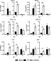
SARS-CoV macrodomain enzymatic activity suppresses early cytokine production in vivo. BALB/c mice were infected as described above, lungs were harvested at the indicated times, and transcript levels were determined by RT-qPCR with primers specific for each transcript and normalized to HPRT. n = 4 mice per group/experiment. Data are derived from results from a single experiment, representative of 2 to 3 independent experiments.
FIG 6
FIG 6
The SARS-CoV macrodomain is required but not sufficient to suppress cytokine expression in vitro. (A) Calu-3 2B4 cells were infected with WT or N1040A virus at an MOI of 2 PFU/cell. Cells were collected at the indicated time points, and RNA levels were determined by RT-qPCR with primers specific for each transcript and normalized to HPRT. Data are derived from results of 1 experiment representative of 3 independent experiments. (B) Calu-3 2B4 cells were infected as described for panel A, and cells were collected at 48 hpi. Lysates were analyzed by immunoblotting with the indicated antibodies using a Li-COR Odyssey Imager. (C and D) pcDNA3-GFP, pLKO-MERS-CoV ORF4A, and pcDNA3-sMacro were transfected into HeLa cells. At 24 h later, the cells were either collected for immunoblotting (C) or transfected with 1 μg pI-C or 1 μg dA-dT (D). For panel D, cells were collected at 16 h after transfection and RNA was analyzed by RT-qPCR with primers specific for indicated transcript. HA, hemagglutinin; GAPDH, glyceraldehyde-3-phosphate dehydrogenase. Data are derived from results of 1 experiment representative of 2 independent experiments. *, P ≤ 0.05; **, P ≤ 0.01; ***, P ≤ 0.001; #, P = 0.06; n.s., not significant.
FIG 7
FIG 7
Mice coinfected with WT and N1040A virus have better outcomes than mice infected with WT virus alone. (A) BALB/c mice were infected with 3 × 10⁴ PFU of WT virus alone or coinfected with 3 × 10⁴ PFU of both WT and N1040A virus. Survival was monitored for 10 days. n = 15 mice for each group. (B) BALB/c mice were infected with 3 × 10⁴ PFU of WT virus or N1040A virus or coinfected with 3 × 10⁴ PFU of both WT and N1040A as described, lungs were harvested at the indicated times, and indicated transcript levels were determined by RT-qPCR with primers specific for each individual gene and then were normalized to HPRT. n = 4 mice per group. (C) BALB/c mice were infected as described for panel B, and virus and lung titers were determined by plaque assay. n = 3 to 4 mice per group. Data are derived from results of 1 experiment representative of 2 independent experiments. *, P ≤ 0.05; **, P ≤ 0.01; ***, P ≤ 0.001; n.s., not significant.

References

    1. Kuny CV, Sullivan CS. 2016. Virus-host interactions and the ARTD/PARP family of enzymes. PLoS Pathog 12:e1005453. doi:10.1371/journal.ppat.1005453. - DOI - PMC - PubMed
    1. Atasheva S, Akhrymuk M, Frolova EI, Frolov I. 2012. New PARP gene with an anti-alphavirus function. J Virol 86:8147–8160. doi:10.1128/JVI.00733-12. - DOI - PMC - PubMed
    1. Goodier JL, Pereira GC, Cheung LE, Rose RJ, Kazazian HH Jr. 2015. The broad-spectrum antiviral protein ZAP restricts human retrotransposition. PLoS Genet 11:e1005252. doi:10.1371/journal.pgen.1005252. - DOI - PMC - PubMed
    1. Leung A, Todorova T, Ando Y, Chang P. 2012. Poly(ADP-ribose) regulates post-transcriptional gene regulation in the cytoplasm. RNA Biol 9:542–548. doi:10.4161/rna.19899. - DOI - PMC - PubMed
    1. Seo GJ, Kincaid RP, Phanaksri T, Burke JM, Pare JM, Cox JE, Hsiang TY, Krug RM, Sullivan CS. 2013. Reciprocal inhibition between intracellular antiviral signaling and the RNAi machinery in mammalian cells. Cell Host Microbe 14:435–445. doi:10.1016/j.chom.2013.09.002. - DOI - PMC - PubMed

Publication types

MeSH terms

LinkOut - more resources