Review
. 2016 Nov 28:7:573.
doi: 10.3389/fphys.2016.00573. eCollection 2016.

Fatty Acid Regulation of Voltage- and Ligand-Gated Ion Channel Function

Affiliations
Review

Fatty Acid Regulation of Voltage- and Ligand-Gated Ion Channel Function

Silvia S Antollini et al. Front Physiol. .

Abstract

Free fatty acids (FFA) are essential components of the cell, where they play a key role in lipid and carbohydrate metabolism, and most particularly in cell membranes, where they are central actors in shaping the physicochemical properties of the lipid bilayer and the cellular adaptation to the environment. FFA are continuously being produced and degraded, and a feedback regulatory function has been attributed to their turnover. The massive increase observed under some pathological conditions, especially in brain, has been interpreted as a protective mechanism possibly operative on ion channels, which in some cases is of stimulatory nature and in other cases inhibitory. Here we discuss the correlation between the structure of FFA and their ability to modulate protein function, evaluating the influence of saturation/unsaturation, number of double bonds, and cis vs. trans isomerism. We further focus on the mechanisms of FFA modulation operating on voltage-gated and ligand-gated ion channel function, contrasting the still conflicting evidence on direct vs. indirect mechanisms of action.

Keywords: FFA; PUFA; VLCFA; cell-surface receptors; fatty acids; ion channels; ligand-gated channel.


Figures
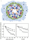
Figure 1
Figure 1
(A) Schematic diagram of a direct mechanism of action of FFA on nAChR function. The scheme illustrates the nAChR-lipid relationship in a receptor-rich membrane, outlining the spatial distribution of the transmembrane segments (TM1, TM2, TM3, and TM4) and the surrounding lipid shell. Three lipid topologies are indicated: non-annular lipids (bordeaux •), annular lipids (green •) and bulk lipids (light blue •). FFA (blue ▴) can be found in any of these three domains. (B,C) Experimental studies on annular and non-annular sites using the efficiency (E) of the Förster resonance energy transfer (FRET) process between the intrinsic fluorescence of T. californica nAChR membranes and the extrinsic fluorescent probe Laurdan; (B) in the presence of increasing concentrations of DOPC (■), cholesterol hemisuccinate (CHS, ▴), and oleic acid (•), where the symbol (♢) corresponds to the sum of E of DOPC and CHS (from Antollini and Barrantes, 1998) and (C) in the presence of increasing concentrations of arachidonic acid with the nAChR in the resting (“R”, ♢) or the desensitized (“D”, ♦) state. The latter was generated by incubation of the membrane with 1 mM carbamoylcholine prior to the fluorescence measurements (From Fernández Nievas et al., 2007).
Figure 2
Figure 2
Sequential model of the nAChR activation equilibrium. The model considers that each state of channel activation has a corresponding desensitized state (Dilger and Liu, 1992): R is the nAChR in the resting state, A the agonist and RA and RA2 represent the nAChR with one or two agonist molecules bound, respectively, and RA2* is the biliganded nAChR in a transient active open configuration; D, AD, A2D, and A2D* are the corresponding isoforms in the desensitized, non-conductive states, respectively. This allosteric equilibrium can be affected e.g., by single-point mutations and exposure to some drugs (From Fernández Nievas et al., 2008).
Figure 3
Figure 3
Schematic diagram of indirect mechanisms of action of FFA on neuronal nAChR function. Different FFA, or their derivatives, can modulate α7 nAChR through various signaling pathways. See text for details.

References

    1. An R. H., Wang X. L., Kerem B., Benhorin J., Medina A., Goldmit M., et al. . (1998). Novel LQT-3 mutation affects Na+ channel activity through interactions between alpha- and beta1-subunits. Circ. Res. 83, 141–146. 10.1161/01.RES.83.2.141 - DOI - PubMed
    1. Anderson M. P., Welsh M. J. (1990). Fatty acid inhibit apical membrane chloride channels in airway epithelia. Proc. Natl. Acad. Sci. U.S.A. 87, 7334–7338. 10.1073/pnas.87.18.7334 - DOI - PMC - PubMed
    1. Andreasen T. J., Doerge D. R., McNamee M. G. (1979). Effects of phospholipase A2 on the binding and ion permeability control properties of the acetylcholine receptor. Arch. Biochem. Biophys. 194, 468–480. 10.1016/0003-9861(79)90641-6 - DOI - PubMed
    1. Andreasen T. J., McNamee M. G. (1977). Phospholipase A inhibition of acetylcholine receptor function in Torpedo californica membrane vesicles. Biochem. Biophys. Res. Commun. 79, 958–965. 10.1016/0006-291X(77)91203-7 - DOI - PubMed
    1. Andreasen T. J., McNamee M. G. (1980). Inhibition of ion permeability control properties of acetylcholine receptor from Torpedo californica by long-chain fatty acids. Biochemistry 30, 4719–4726. 10.1021/bi00561a027 - DOI - PubMed

LinkOut - more resources