

Killian-Jamieson diverticulum: real-time sonographic findings
- PMID: 27965721
- PMCID: PMC5126011
- DOI: 10.1007/s40477-016-0208-3
Killian-Jamieson diverticulum: real-time sonographic findings
Abstract
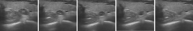
Killian-Jamieson diverticulum (KJD) is a pharyngoesophageal diverticulum that can be observed during a neck ultrasound examination. Because of its position, it is frequently misinterpreted as a thyroid nodule. We present a case of an incidental finding of KJD, where changes in shape during dynamic scanning led to the correct diagnosis, preventing from invasive procedures such as fine needle aspiration.
Il diverticolo di Killian–Jamieson (KJD) è un diverticolo faringoesofageo che può essere individuato durante una valutazione ultrasonografica del collo. A causa della sua posizione, è frequentemente interpretato come un nodulo tiroideo. Presentiamo un caso di un riscontro occasionale di KJD, in cui le variazioni della forma durante le scansioni dinamiche hanno condotto ad una corretta diagnosi, evitando procedure invasive quali il prelievo con agoaspirato.
Conflict of interest statement
The authors have no conflict of interest. Informed consent All procedures followed were in accordance with the ethical standards of the responsible committee on human experimentation (institutional and national) and with the Helsinki Declaration of 1975, as revised in 2000. All patients provided written informed consent for enrolment in the study and for the inclusion in this article of information that could potentially lead to their identification. Human and animal studies The study was conducted in accordance with all institutional and national guidelines for the care and use of laboratory animals.
Figures


References
-
- Killian G. Über den Mund der Speiseröhre. Zeitschrift für Ohrenheilkunde. 1908;55:1–44.
-
- Jamieson EB (1934) Illustrations of regional anatomy. E&S Livingstone Ltd, Edinburgh, p 44
Publication types
MeSH terms
LinkOut - more resources
Full Text Sources
Other Literature Sources
