Review
. 2016 Nov 29:4:140.
doi: 10.3389/fcell.2016.00140. eCollection 2016.

Diacylglycerol Kinases: Shaping Diacylglycerol and Phosphatidic Acid Gradients to Control Cell Polarity

Affiliations
Review

Diacylglycerol Kinases: Shaping Diacylglycerol and Phosphatidic Acid Gradients to Control Cell Polarity

Gianluca Baldanzi et al. Front Cell Dev Biol. .

Abstract

Diacylglycerol kinases (DGKs) terminate diacylglycerol (DAG) signaling and promote phosphatidic acid (PA) production. Isoform specific regulation of DGKs activity and localization allows DGKs to shape the DAG and PA gradients. The capacity of DGKs to constrain the areas of DAG signaling is exemplified by their role in defining the contact interface between T cells and antigen presenting cells: the immune synapse. Upon T cell receptor engagement, both DGK α and ζ metabolize DAG at the immune synapse thus constraining DAG signaling. Interestingly, their activity and localization are not fully redundant because DGKζ activity metabolizes the bulk of DAG in the cell, whereas DGKα limits the DAG signaling area localizing specifically at the periphery of the immune synapse. When DGKs terminate DAG signaling, the local PA production defines a new signaling domain, where PA recruits and activates a second wave of effector proteins. The best-characterized example is the role of DGKs in protrusion elongation and cell migration. Indeed, upon growth factor stimulation, several DGK isoforms, such as α, ζ, and γ, are recruited and activated at the plasma membrane. Here, local PA production controls cell migration by finely modulating cytoskeletal remodeling and integrin recycling. Interestingly, DGK-produced PA also controls the localization and activity of key players in cell polarity such as aPKC, Par3, and integrin β1. Thus, T cell polarization and directional migration may be just two instances of the general contribution of DGKs to the definition of cell polarity by local specification of membrane identity signaling.

Keywords: cell polarity; immune synapse; lipid domain; localization; migration.


Figures

Figure 1
Figure 1
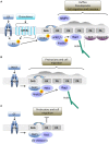
The role of DGKα in shaping diacylglycerol signaling at the immune synapse. Upon TCR/SLAM engagement, PLCγ activation induces DAG production at the synapse and DGKα/ζ recruitment. DGKζ (blue dotted line) is responsible for the metabolism of the bulk of DAG over the entire synapse, whereas DGKα (light orange) is recruited by PI3Kδ generated PI3,4,5P3 at the pSMAC, where DAG is constrained. Excessive DAG metabolism at the IS by DGKα is prevented by SAP-mediated inhibition. The resulting sharp DAG gradient promotes downstream MAPK signaling and the local recruitment of nPKC, which promotes MTOC reorientation.
Figure 2
Figure 2
DGKs-produced PA drives membrane protrusion and cell motility. (A) In epithelial cells, growth factors or chemokine stimulation promotes Src mediated recruitment and activation of DGKα to nascent protrusions. The resulting PA enriched domain drives protrusion elongation and matrix invasion by the local recruitment of aPKC and Rab11-FIP1. These PA binding proteins respectively control cytoskeletal remodeling through Rac1 and integrin β1 recycling. (B) In fibroblasts, PDGF triggers DGKζ recruitment to nascent protrusions. The resulting PA enriched domain drives focal adhesion remodeling and cell migration by recruiting and activating PAK1 and promoting Rac1 activation. (C) Growth factors also promote membrane recruitment and activation of DGKγ that limits Rac1 activity through β2 chimaerin.

References

    1. Abramovici H., Mojtabaie P., Parks R. J., Zhong X. P., Koretzky G. A., Topham M. K., et al. . (2009). Diacylglycerol kinase zeta regulates actin cytoskeleton reorganization through dissociation of Rac1 from RhoGDI. Mol. Biol. Cell 20, 2049–2059. 10.1091/mbc.E07-12-1248 - DOI - PMC - PubMed
    1. Almena M., Mérida I. (2011). Shaping up the membrane: diacylglycerol coordinates spatial orientation of signaling. Trends Biochem. Sci. 36, 593–603. 10.1016/j.tibs.2011.06.005 - DOI - PubMed
    1. Alonso R., Mazzeo C., Rodriguez M. C., Marsh M., Fraile-Ramos A., Calvo V., et al. . (2011). Diacylglycerol kinase α regulates the formation and polarisation of mature multivesicular bodies involved in the secretion of Fas ligand-containing exosomes in T lymphocytes. Cell Death Differ. 18, 1161–1173. 10.1038/cdd.2010.184 - DOI - PMC - PubMed
    1. Ard R., Mulatz K., Abramovici H., Maillet J. C., Fottinger A., Foley T., et al. . (2012). Diacylglycerol kinase ζ regulates RhoA activation via a kinase-independent scaffolding mechanism. Mol. Biol. Cell 23, 4008–4019. 10.1091/mbc.E12-01-0026 - DOI - PMC - PubMed
    1. Avila-Flores A., Santos T., Rincón E., Mérida I. (2005). Modulation of the mammalian target of rapamycin pathway by diacylglycerol kinase-produced phosphatidic acid. J. Biol. Chem. 280, 10091–10099. 10.1074/jbc.M412296200 - DOI - PubMed