IL-6 Promotes Islet β-Cell Dysfunction in Rat Collagen-Induced Arthritis
- PMID: 27965984
- PMCID: PMC5124658
- DOI: 10.1155/2016/7592931
IL-6 Promotes Islet β-Cell Dysfunction in Rat Collagen-Induced Arthritis
Abstract
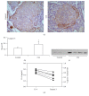
The aim of this study was to explore the possible mechanism of rheumatoid arthritis- (RA-) related abnormal glucose metabolism. The model of collagen-induced arthritis (CIA) was established by intradermal injection of type II collagen into Wistar rats; complete Freund's adjuvant injections were used as the control group. Fasting plasma glucose (FBG) was measured by the glucose oxidase method. Fasting insulin (FIns) and the expressions of IL-6 were detected by ELISA. Islet caspase-3 was examined by immunohistochemistry. On day 17 after immunization, FBG of the CIA group showed an elevated FBG value compared with the control group. Meanwhile, the FIns of group CIA was lower when compared with the control group. Interestingly, the inflammatory cytokine IL-6 expression was significantly increased when compared with the control group. As expected, the abnormal glucose metabolism was accompanied by the increased IL-6 expression. Furthermore, in line with the upregulated IL-6 expression, the apoptosis related enzyme caspase-3 was also markedly increased. These data showed that the elevated FBG in CIA may be associated with the reduced FIns level secondary to the overapoptosis of pancreas islet cells induced by IL-6.
Conflict of interest statement
The authors declare no competing interests regarding the publication of this paper.
Figures


Similar articles
-
Abnormal Glucose Metabolism in Rheumatoid Arthritis.Biomed Res Int. 2017;2017:9670434. doi: 10.1155/2017/9670434. Epub 2017 Apr 26. Biomed Res Int. 2017. PMID: 28529957 Free PMC article. Review.
-
Extract of the dried heartwood of Caesalpinia sappan L. attenuates collagen-induced arthritis.J Ethnopharmacol. 2011 Jun 14;136(1):271-8. doi: 10.1016/j.jep.2011.04.061. Epub 2011 Apr 30. J Ethnopharmacol. 2011. PMID: 21557995
-
Early deficits in insulin secretion, beta cell mass and islet blood perfusion precede onset of autoimmune type 1 diabetes in BioBreeding rats.Diabetologia. 2018 Apr;61(4):896-905. doi: 10.1007/s00125-017-4512-z. Epub 2017 Dec 6. Diabetologia. 2018. PMID: 29209740 Free PMC article.
-
Beta-endorphin prevents collagen induced arthritis by neuroimmuno-regulation pathway.Neuro Endocrinol Lett. 2005 Dec;26(6):739-44. Neuro Endocrinol Lett. 2005. PMID: 16380673
-
Protection against cartilage and bone destruction by systemic interleukin-4 treatment in established murine type II collagen-induced arthritis.Arthritis Res. 1999;1(1):81-91. doi: 10.1186/ar14. Epub 1999 Oct 26. Arthritis Res. 1999. PMID: 11056663 Free PMC article.
Cited by
-
Network Pharmacology-Based Strategy for Exploring the Pharmacological Mechanism of Honeysuckle (Lonicer japonica Thunb.) against Newcastle Disease.Evid Based Complement Alternat Med. 2022 Apr 5;2022:9265094. doi: 10.1155/2022/9265094. eCollection 2022. Evid Based Complement Alternat Med. 2022. PMID: 35422871 Free PMC article.
-
Abnormal Glucose Metabolism in Rheumatoid Arthritis.Biomed Res Int. 2017;2017:9670434. doi: 10.1155/2017/9670434. Epub 2017 Apr 26. Biomed Res Int. 2017. PMID: 28529957 Free PMC article. Review.
References
MeSH terms
Substances
LinkOut - more resources
Full Text Sources
Other Literature Sources
Medical
Research Materials

