. 2017 Jan 20;28(3):035101.
doi: 10.1088/1361-6528/28/3/035101. Epub 2016 Dec 14.

Optical properties of biomimetic probes engineered from erythrocytes

Affiliations

Optical properties of biomimetic probes engineered from erythrocytes

Joshua M Burns et al. Nanotechnology. .

Abstract

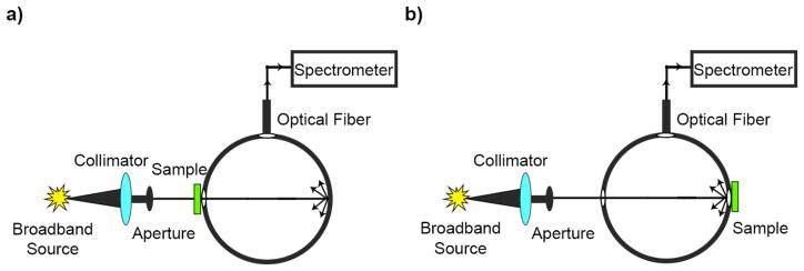
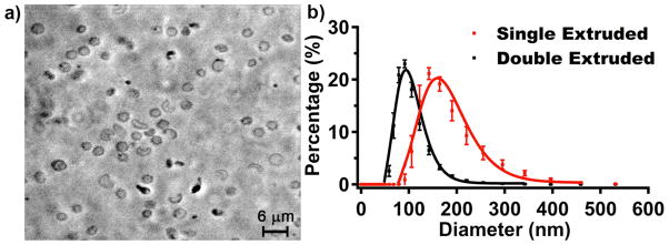
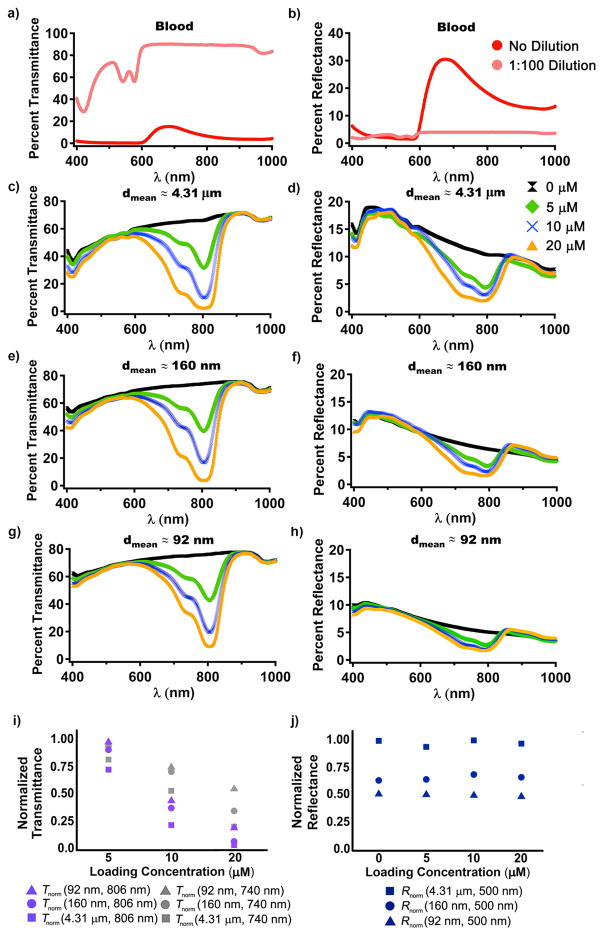
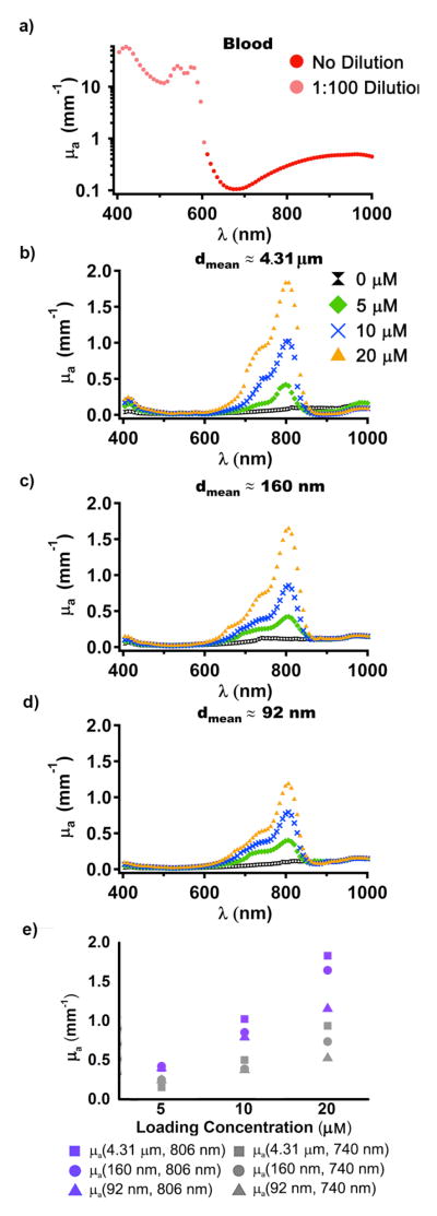
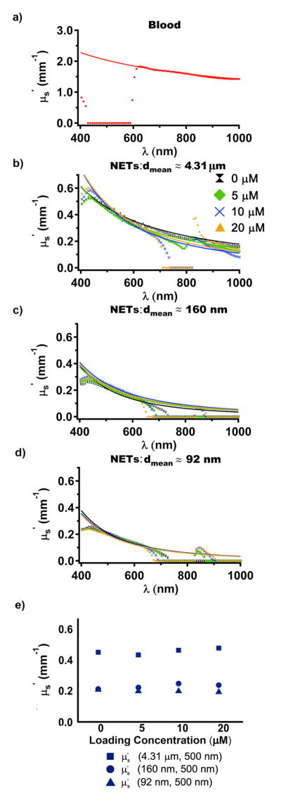
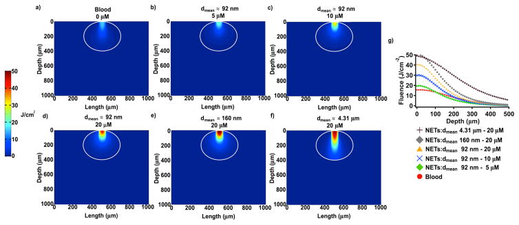
Light-activated theranostic materials offer a potential platform for optical imaging and phototherapeutic applications. We have engineered constructs derived from erythrocytes, which can be doped with the FDA-approved near infrared (NIR) chromophore, indocyanine green (ICG). We refer to these constructs as NIR erythrocyte-mimicking transducers (NETs). Herein, we investigated the effects of changing the NETs mean diameter from micron- (≈4 μm) to nano- (≈90 nm) scale, and the ICG concentration utilized in the fabrication of NETs from 5 to 20 μM on the resulting absorption and scattering characteristics of the NETs. Our approach consisted of integrating sphere-based measurements of light transmittance and reflectance, and subsequent utilization of these measurements in an inverse adding-doubling algorithm to estimate the absorption (μ a) and reduced scattering (μ s') coefficients of these NETs. For a given NETs diameter, values of μ a increased over the approximate spectral band of 630-860 nm with increasing ICG concentration. Micron-sized NETs produced the highest peak value of μ a when using ICG concentrations of 10 and 20 μM, and showed increased values of μ s' as compared to nano-sized NETs. Spectral profiles of μ s' for these NETs showed a trend consistent with Mie scattering behavior for spherical objects. For all NETs investigated, changing the ICG concentration minimally affected the scattering characteristics. A Monte Carlo-based model of light distribution showed that the presence of these NETs enhanced the fluence levels within simulated blood vessels. These results provide important data towards determining the appropriate light dosimetry parameters for an intended light-based biomedical application of NETs.


Figures

Figure 1
Figure 1
Schematic of integrating sphere-based measurements of (a) transmittance and (b) reflectance for whole blood, EGs, and NET samples.
Figure 2
Figure 2
Phase contrast image of micron-sized NETs (a) formed without extrusion, and hydrodynamic diameter distributions of nano-sized NETs (b) formed by single or double extrusions, as determined by DLS method. The respective ranges of the measured diameters, based on DLS, for NETs fabricated by single and double extrusion methods were ≈ 91–396 nm, and 59–190 nm. We present the mean and standard deviation of the measurements of these samples (n = 6), represented as circles and error bars, respectively. The estimated mean diameters as determined from the lognormal fits (solid curves) were ≈ 160 nm and 92 nm for NETs formed by single and double extrusion methods, respectively.
Figure 3
Figure 3
Spectrally dependent values of percent transmittance (a), and reflectance (b) for whole bovine blood samples without dilution, and diluted 100 times by adding 1x PBS (≈310 mOsm). Spectra of percent transmittance (c, e, g), and reflectance (d, f, h) for micron-sized NETs formed without extrusion (c, d), or by single (e, f) or double extrusions (g, h) using various ICG concentrations. Measurements were obtained with NETs suspended in 1x PBS (≈ 310 mOsm). The legend labels indicate the ICG concentration levels in the loading buffer. In panel (i), transmittance measurements are normalized to the values at 740 nm and 806 nm for 92 nm NETs formed using 5 μM ICG in the loading buffer. In panel (j), reflectance measurements are normalized to the value at 500 nm (spectral peak) for EGs.
Figure 4
Figure 4
Spectrally-dependent values of μa for whole bovine blood (a) without dilution for λ > 604 nm, and the sample diluted 100 times by adding 1x PBS (≈ 310 mOsm) for λ ≤ 604 nm. Values of μa for the diluted sample were multiplied by 100. Spectra of μa for micron-sized NETs formed without extrusion (b), or by single (c) or double extrusions (d) using various ICG concentrations in the loading buffer. Measurements were obtained with NETs suspended in 1x PBS (≈ 310 mOsm). In panel (e), we show a summary of μa values at 740 nm and 810 nm.
Figure 5
Figure 5
Spectrally dependent values of μs′ for whole bovine blood (a) without dilution and the fitted profile based on equation (2). Spectra of μs′ for micron-sized NETs formed without extrusion (b), or by single (c) or double extrusions (d) using various ICG concentrations in the loading buffer, and the fitted profile based on equation (2). Measurements were obtained with NETs suspended in 1x PBS (≈ 310 mOsm). In panel (e), we show a summary of μs′ values at 500 nm.
Figure 6
Figure 6
Monte Carlo-based estimations of fluence profiles within a simulated 500 μm diameter blood vessel filled with blood containing 45% hematocrit without added NETs (a), or with added nano-sized NETs (dmean ≈ 92 nm) (b–d), dmean ≈ 160 nm NETs (c), micron-sized NETS (dmean ≈ 4.31 μm). Relative fractions of blood and NETs in panels (b–e) were 50%. We utilized the optical properties of NETs formed using ICG concentrations of 5, 10, and 20 μM (b–d), and 20 μM (e, f) in the loading buffer. We assumed 806 nm laser focused on a 100 μm diameter spot at incident fluence of 10 J/cm2. Color scale bar represents the resulting fluence in J/cm2 and white circle represents outline of the blood vessel. The white margins delineate the boundaries of the blood vessels. In panel (g), we show the depth profile of the fluences along the central axis of the laser beam.

Similar articles

Cited by

References

    1. Kim H, Chung K, Lee S, Kim DH, Lee H. Near-infrared light-responsive nano-materials for cancer theranostics. WIREs Nanomed Nanobiotechnol. 2016;8:23–25. - PubMed
    1. Cheng L, Yuan C, Shen S, Yi X, Gong H, Yang K, Liu Z. Bottom-up synthesis of metal-ion-doped WS2 nanoflakes for cancer theranostics. ACS Nano. 2015;9:11090–11101. - PubMed
    1. Shan G, Weissleder R, Hilderbrand SA. Upconverting organic dye doped core-shell nano-composites for dual-modality NIR imaging and photo-thermal therapy. Theranostics. 2013;3:267–274. - PMC - PubMed
    1. Lee M-Y, Lee C, Jung HS, Jeon M, Kim KS, Yun SH, Kim C, Hahn SK. Biodegradable photonic melanoidin for theranostic applications. ACS Nano. 2016;10:822–831. - PubMed
    1. Lin L-S, Yang X, Niu G, Song J, Yang H-H, Chen X. Dual-enhanced photothermal conversion properties of reduced graphene oxide-coated gold superparticles for light-triggered acoustic and thermal theranostics. Nanoscale. 2016;8:2116–2122. - PMC - PubMed