. 2016 Dec 14:6:39048.
doi: 10.1038/srep39048.

Molecular mechanism: the human dopamine transporter histidine 547 regulates basal and HIV-1 Tat protein-inhibited dopamine transport

Affiliations

Molecular mechanism: the human dopamine transporter histidine 547 regulates basal and HIV-1 Tat protein-inhibited dopamine transport

Pamela M Quizon et al. Sci Rep. .

Abstract

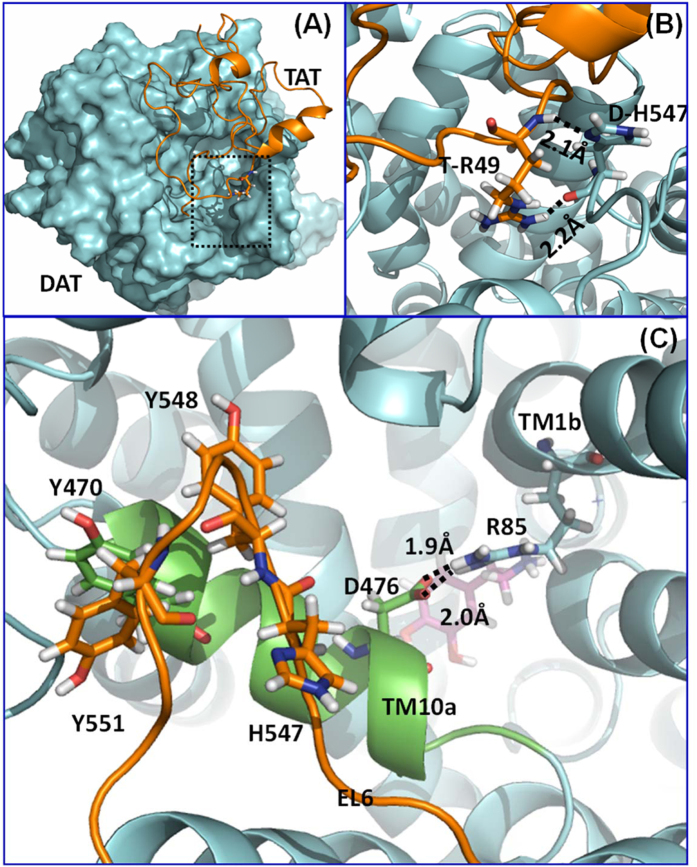
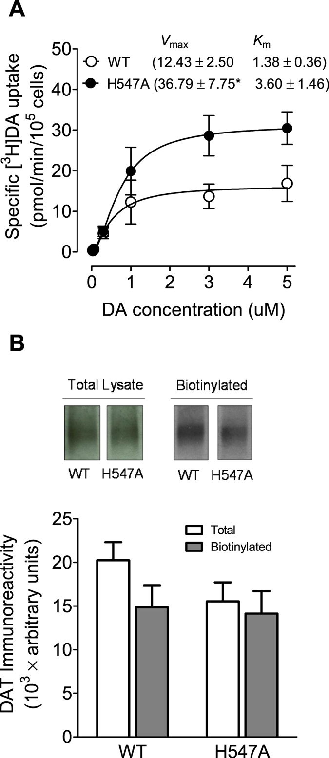
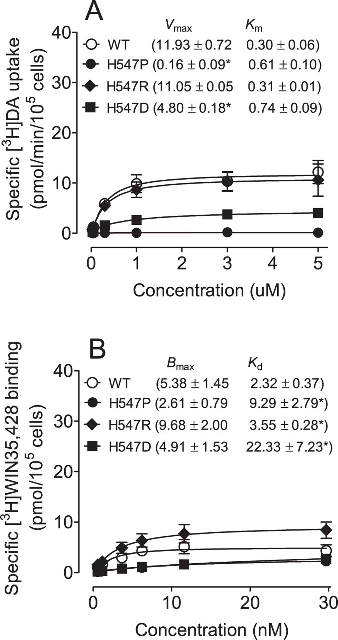
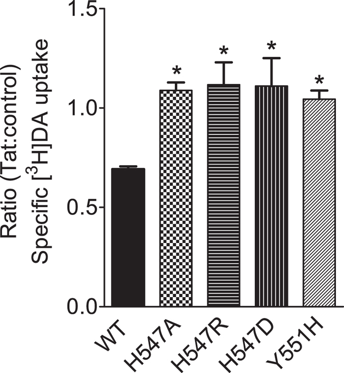
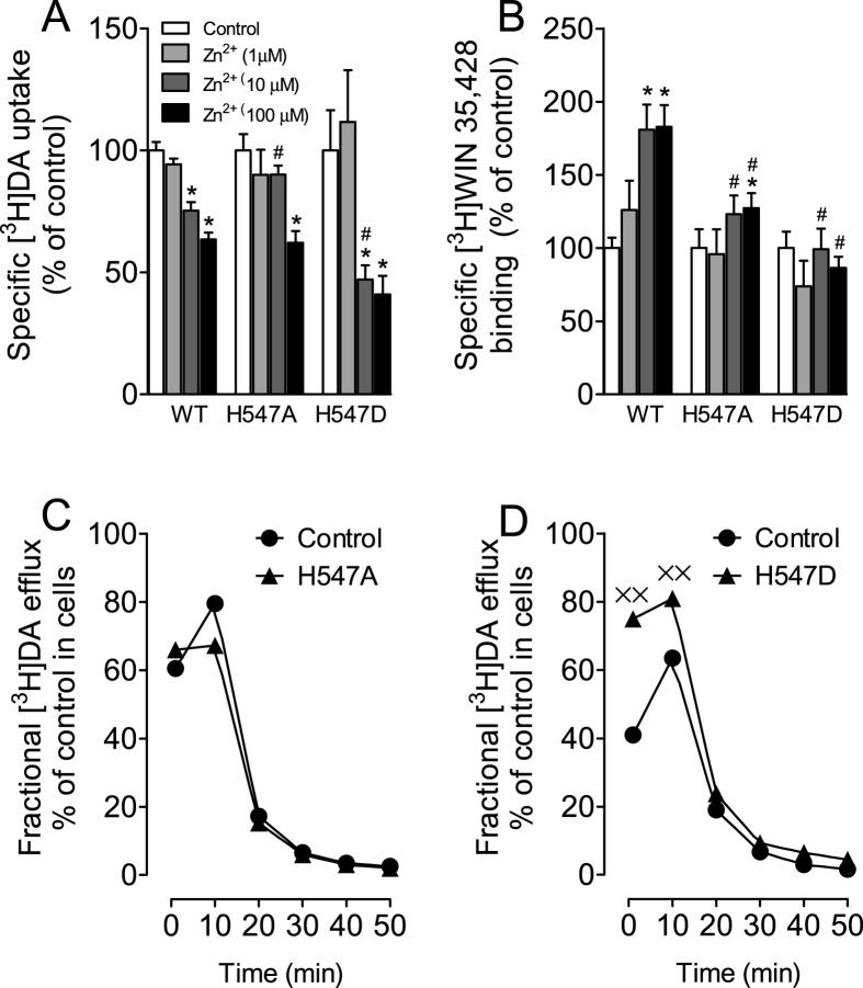
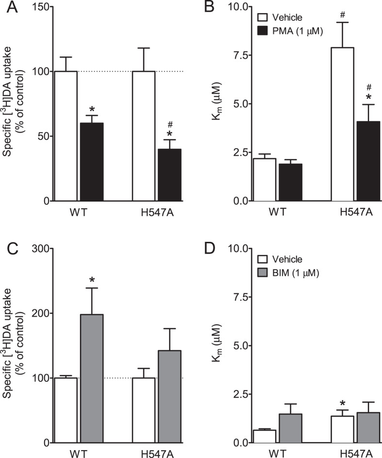
Abnormal dopaminergic transmission has been implicated as a risk determinant of HIV-1-associated neurocognitive disorders. HIV-1 Tat protein increases synaptic dopamine (DA) levels by directly inhibiting DA transporter (DAT) activity, ultimately leading to dopaminergic neuron damage. Through integrated computational modeling prediction and experimental validation, we identified that histidine547 on human DAT (hDAT) is critical for regulation of basal DA uptake and Tat-induced inhibition of DA transport. Compared to wild type hDAT (WT hDAT), mutation of histidine547 (H547A) displayed a 196% increase in DA uptake. Other substitutions of histidine547 showed that DA uptake was not altered in H547R but decreased by 99% in H547P and 60% in H547D, respectively. These mutants did not alter DAT surface expression or surface DAT binding sites. H547 mutants attenuated Tat-induced inhibition of DA transport observed in WT hDAT. H547A displays a differential sensitivity to PMA- or BIM-induced activation or inhibition of DAT function relative to WT hDAT, indicating a change in basal PKC activity in H547A. These findings demonstrate that histidine547 on hDAT plays a crucial role in stabilizing basal DA transport and Tat-DAT interaction. This study provides mechanistic insights into identifying targets on DAT for Tat binding and improving DAT-mediated dysfunction of DA transmission.


Figures

Figure 1
Figure 1
(A) Computational model of human dopamine transporter (hDAT) and HIV-1 Tat binding. hDAT and Tat are represented as cyan surface and golden ribbon, respectively. (B) A local view of DAT residue H547 and its direct interaction with Tat residue R49, hDAT is represented as a cyan ribbon. The residues are represented in sticks, with hydrogen bonds between the two residues represented with dashed lines labeled with their corresponding coordination distances. (C) Structural details of the residues Y470, Y551, H547, D476, and R85 on hDAT. hDAT is represented as a cyan ribbon, while the first part of transmembrane helix 10 (TM10a) and extracellular loop 6 (EL6) are colored in green and orange, respectively. Dopamine is represented as a ball-and-stick molecule in purple. Hydrogen bonds between D476 and R85 are indicated by dashed lines with coordinating distances labeled.
Figure 2
Figure 2. DA transport and DAT surface expression in WT hDAT and mutant.
(A) Kinetic analysis of [3H]DA uptake in WT hDAT and H547A-hDAT. PC12 cells transfected with WT hDAT or H547A-hDAT were incubated with one of 6 mixed concentrations of [3H]DA as total rate of DA uptake. In parallel, nonspecific uptake of each concentration of [3H]DA (in the presence of 10 μM nomifensine, final concentration) was subtracted from total uptake to calculate DAT-mediated uptake. The Vmax and Km values were estimated by fitting the data to the Michaelis-Menten equation and represent the means from five independent experiments ± S.E.M. *p < 0.05 compared to control value (unpaired Student’s t test) (n = 5). (B) Cell surface expression of WT hDAT and H547A-hDAT was analyzed by biotinylation assay. Top panel: representative immunoblots (see supplemental information) PC12 cells expressing WT hDAT (WT) or H547A-hDAT (H547A) (n = 9).
Figure 3
Figure 3. DA transport and DAT surface binding sites in WT hDAT and H547 substitutional mutants.
(A) Kinetic analysis of [3H]DA uptake in WT hDAT and mutants. The Vmax and Km values were estimated by fitting the data to the Michaelis-Menten equation and represent the means from five independent experiments ± S.E.M. *p < 0.05 compared to WT hDAT value (unpaired Student’s t test) (n = 5). (B) Saturation binding of [3H]WIN35,428 in intact PC12 cells transfected with WT hDAT and mutants. The Bmax and Kd values were estimated by fitting the data on a one-site binding curve and represent the means from four independent experiments ± S.E.M. *p < 0.05 compared to control value (unpaired Student’s t test) (n = 4).
Figure 4
Figure 4. Effects of Tat on kinetic analysis of [3H]DA uptake in WT hDAT and His547 mutants.
PC12 cells transfected with WT hDAT (WT), H547A-hDAT (H547A), H547R-hDAT (H547R), H547D-hDAT (H547D), or Y551H-hDAT (Y551H) were preincubated with or without recombinant Tat1–86 (rTat1–86) (140 nM, final concentration) at room temperature for 20 min followed by the addition of [3H]DA. Nonspecific uptake was determined in the presence of 10 μM final concentration of nomifensine. Data are expressed as the ratio of the specific [3H]DA uptake in the presence of Tat to that in the absence of Tat [in DPM: WT hDAT (Tat, 4794 ± 989 vs control, 6858 ± 1393); H547A (Tat, 6190 ± 1474 vs control, 5799 ± 1408); H547R (Tat, 5026 ± 1097 vs control, 4698 ± 1257); H547D (Tat, 2159 ± 557 vs control, 2021 ± 565); and Y551H (Tat, 662 ± 107 vs control, 642 ± 108)] n = 7–8. *p < 0.05 compared with WT hDAT control values.
Figure 5
Figure 5. Effects of H547A and H547D mutants on transporter conformational transitions.
Mutations of His547 affect zinc regulation of [3H]DA uptake (A) and [3H]WIN 35,428 binding (B). PC12 cells transfected with WT hDAT (WT), H547A-hDAT (H547A) and H547D-hDAT (H547D) were incubated with KRH buffer alone (control) or ZnCl2 (1, 10, 100 μM, final concentration) followed by [3H]DA uptake or [3H]WIN 35,428 binding (n = 5–7). The histogram shows [3H]DA uptake and [3H]WIN 35,428 binding expressed as mean ± S.E.M. of the respective controls set to 100% for the mutant. *p < 0.05 compared to control. #p < 0.05 compared to WT hDAT with ZnCl2. Functional DA efflux of DA properties of H547A-hDAT (C) and H547D-hDAT (D) with their respective WT hDAT control. PC12 cells transfected with WT hDAT or mutants were preincubated with KRH buffer containing [3H]DA (0.05 μM, final concentration) at room temperature for 20 min. After incubation, cells were washed and incubated with fresh buffer as indicated time points. Subsequently, the buffer was removed from cells, and radioactivity in the buffer and remaining in the cells was counted. Each fractional efflux of [3H]DA in WT hDAT (WT) or mutants was expressed as percentage of total [3H]DA in the cells at the start of the experiment. Fractional [3H]DA efflux levels at 1, 10, 20, 30, 40 and 50 min are expressed as the percentage of total [3H]DA with preloading with 0.05 μM (WT hDAT: 13743 ± 3050 dpm, H547A-hDAT: 14464 ± 2547 dpm and H547D-hDAT: 1891 ± 428 dpm) presented in the cells at the start of the experiment (n = 4). ××p < 0.05 compared to WT hDAT (Bonferroni t-test).
Figure 6
Figure 6. Effects of H547A on basal PKC-mediated regulation of DAT function.
Kinetic analysis of [3H]DA uptake in PC12 cells transfected with WT hDAT or H547A-hDAT in the presence or absence of PKC activator PMA (1 μM) (A,B) or inhibitor BIM (1 μM) (C,D). *p < 0.05 compared to their respective controls. #p < 0.05 compared to WT hDAT (n = 6–9).

References

    1. Heaton R. K. et al.. HIV-associated neurocognitive disorders persist in the era of potent antiretroviral therapy: CHARTER Study. Neurology 75, 2087–2096 (2010). - PMC - PubMed
    1. McArthur J. C., Steiner J., Sacktor N. & Nath A. Human immunodeficiency virus-associated neurocognitive disorders: Mind the gap. Ann Neurol 67, 699–714 (2010). - PubMed
    1. King J. E., Eugenin E. A., Buckner C. M. & Berman J. W. HIV tat and neurotoxicity. Microbes and infection/Institut Pasteur 8, 1347–1357 (2006). - PubMed
    1. Clifford D. B. & Ances B. M. HIV-associated neurocognitive disorder. The Lancet infectious diseases 13, 976–986 (2013). - PMC - PubMed
    1. Wang G. J. et al.. Decreased brain dopaminergic transporters in HIV-associated dementia patients. Brain 127, 2452–2458 (2004). - PubMed

Publication types

Substances

LinkOut - more resources