. 2017 Apr 15;195(8):1000-1009.
doi: 10.1164/rccm.201604-0822OC.

Matrix Metalloproteinase-1 Activation Contributes to Airway Smooth Muscle Growth and Asthma Severity

Affiliations

Matrix Metalloproteinase-1 Activation Contributes to Airway Smooth Muscle Growth and Asthma Severity

Shams-Un-Nisa Naveed et al. Am J Respir Crit Care Med. .

Abstract

Rationale: Matrix metalloproteinase-1 (MMP-1) and mast cells are present in the airways of people with asthma.

Objectives: To investigate whether MMP-1 could be activated by mast cells and increase asthma severity.

Methods: Patients with stable asthma and healthy control subjects underwent spirometry, methacholine challenge, and bronchoscopy, and their airway smooth muscle cells were grown in culture. A second asthma group and control subjects had symptom scores, spirometry, and bronchoalveolar lavage before and after rhinovirus-induced asthma exacerbations. Extracellular matrix was prepared from decellularized airway smooth muscle cultures. MMP-1 protein and activity were assessed.

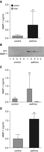
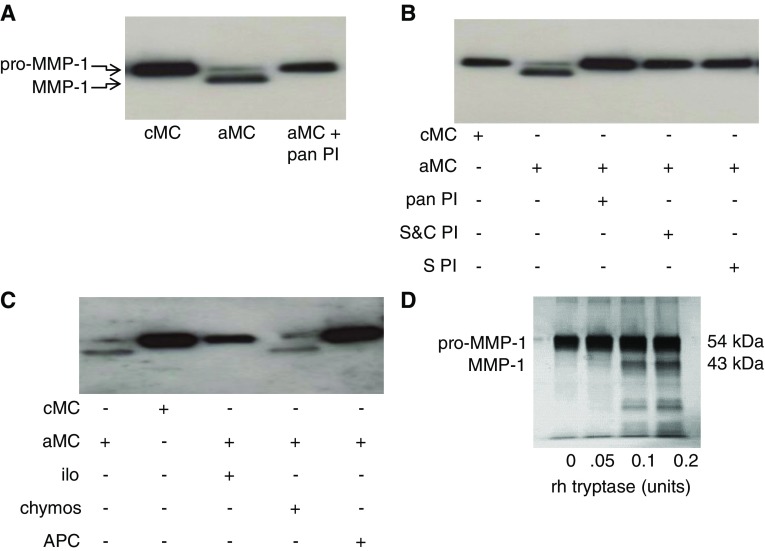
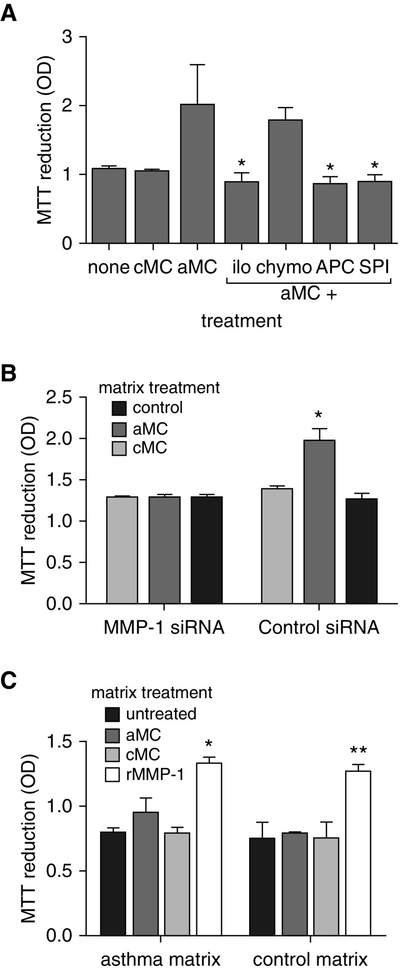
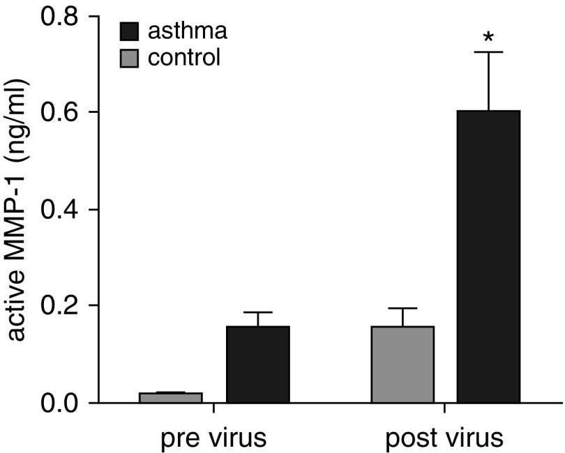
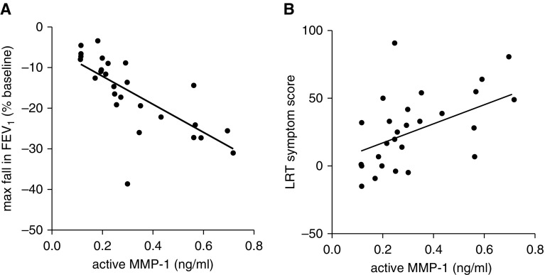
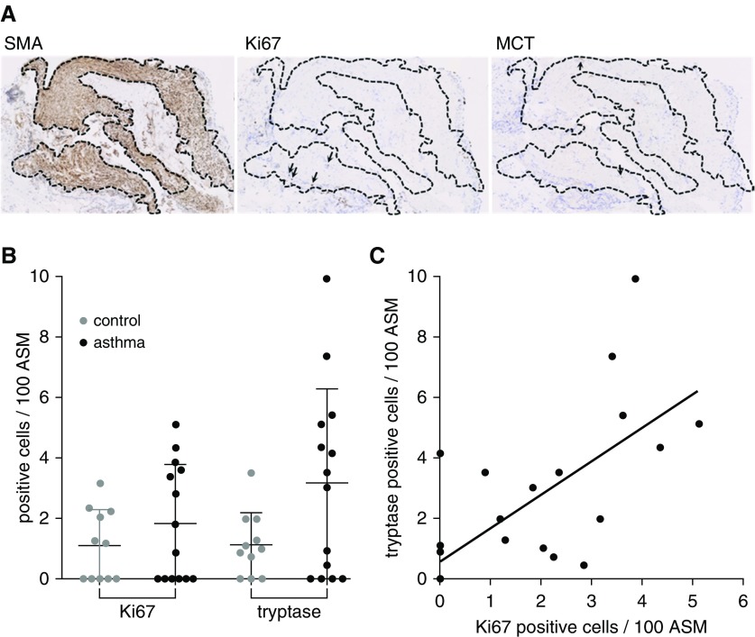
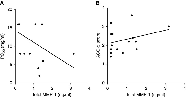
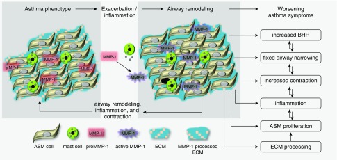
Measurements and main results: Airway smooth muscle cells generated pro-MMP-1, which was proteolytically activated by mast cell tryptase. Airway smooth muscle treated with activated mast cell supernatants produced extracellular matrix, which enhanced subsequent airway smooth muscle growth by 1.5-fold (P < 0.05), which was dependent on MMP-1 activation. In asthma, airway pro-MMP-1 was 5.4-fold higher than control subjects (P = 0.002). Mast cell numbers were associated with airway smooth muscle proliferation and MMP-1 protein associated with bronchial hyperresponsiveness. During exacerbations, MMP-1 activity increased and was associated with fall in FEV1 and worsening asthma symptoms.

Conclusions: MMP-1 is activated by mast cell tryptase resulting in a proproliferative extracellular matrix. In asthma, mast cells are associated with airway smooth muscle growth, MMP-1 levels are associated with bronchial hyperresponsiveness, and MMP-1 activation are associated with exacerbation severity. Our findings suggest that airway smooth muscle/mast cell interactions contribute to asthma severity by transiently increasing MMP activation, airway smooth muscle growth, and airway responsiveness.

Keywords: airway remodeling; airway smooth muscle; asthma; extracellular matrix; mast cells.


Figures

Figure 1.
Figure 1.
Matrix metalloproteinase (MMP)-1 expression in asthma. (A) Total MMP-1 protein measured by ELISA in bronchial washings from 16 patients with mild or moderate asthma, defined by the Global Initiative for Asthma criteria and 11 healthy control subjects. Those with asthma have significantly higher MMP-1 levels. **P = 0.002, Mann-Whitney U test. (B) Western blot of bronchial washings from five representative patients with asthma and five healthy control subjects. A single band of 55 kD consistent with pro–MMP-1 is present in those with asthma but not detectable in healthy control subjects. (C) Total and active MMP-1 activity measured by fluorokine activity assay in patients described in A. Total MMP-1 protein is low in healthy control subjects and significantly elevated in those with asthma, **P < 0.01. MMP-1 activity is undetectable in control subjects and low in those with asthma. (D) Total MMP-1 protein measured by ELISA in cell culture supernatants from airway smooth muscle cells grown from bronchial biopsies from patients with mild or moderate asthma and healthy control subjects. **P = 0.002, Mann-Whitney U test.
Figure 2.
Figure 2.
Mast cell tryptase activates matrix metalloproteinase (MMP)-1. (A) Western blot for MMP-1 in airway smooth muscle supernatants treated with control or activated mast cell supernatants. Activated mast cell supernatants cause MMP-1 activation as shown by the appearance of the smaller MMP-1 band, which is blocked by a broad-spectrum protease inhibitor. (B and C) MMP-1 activation by activated mast cell supernatants is inhibited by inhibitors of serine and cysteine proteases, serine proteases, metalloproteinases, and tryptase, but not chymase. (D) Silver stain of sodium dodecyl sulfate–polyacrylamide gel electrophoresis showing recombinant human tryptase cleaves pro–MMP-1 in vitro shown by the dose-dependent appearance of a 43-kD band. aMC = activated mast cell supernatants; APC = inhibitor of tryptase; chymos = inhibitor of chymase; cMC = control mast cell supernatants; ilo = inhibitor of metalloproteinases; pan PI = broad-spectrum protease inhibitor; rh = recombinant human; S&C PI = inhibitor of serine and cysteine proteases; S PI = inhibitor of serine protease.
Figure 3.
Figure 3.
Mast cell treatment of airway smooth muscle (ASM) generates proproliferative extracellular matrix. ASM from patients with asthma or control subjects was treated with control or activated mast cell supernatants or mast cell activation vehicle during extracellular matrix deposition. ASM cells were removed from extracellular matrix preparations, and normal ASM was seeded for 48 hours in the presence of 1% serum. Cell proliferation was quantitated by (A) 3-(4,5-dimethylthiazol-2-yl)-2,5-diphenyltetrazolium bromide assay (B) and cell counting. ASM cell proliferation was enhanced by activated, but not control, mast cell treatment. **P = 0.0001, two-way analysis of variance. aMC = activated mast cell supernatants; cMC = control mast cell supernatants; MTT = 3-(4,5-dimethylthiazol-2-yl)-2,5-diphenyltetrazolium bromide; OD = optical density.
Figure 4.
Figure 4.
Proproliferative matrix is dependent on airway smooth muscle (ASM)-derived matrix metalloproteinase (MMP)-1. (A) ASM treated with control (cMC) or activated (aMC) mast cell supernatants during extracellular matrix (ECM) deposition was coincubated with an MMP inhibitor, chymase inhibitor, tryptase inhibitor, or a serine protease inhibitor. Enhanced ASM proliferation generated by aMC treatment was abrogated by inhibitors of MMPs, serine proteases, and tryptase but not chymase. *Difference from aMC, P = 0.029, Mann-Whitney U test. (B) ECM preparations were generated by ASM cells treated by an MMP-1–specific or control siRNA. During ECM deposition cells were treated with cMC or aMC. The MMP-1–specific siRNA abrogated the enhanced proliferation generated by aMC treatment. *P = 0.04 Mann-Whitney U test. (C) ASM-derived ECM from asthma-derived or control cells were decellularized and then left untreated or incubated with cMC or aMC or active recombinant MMP-1 (rMMP-1). ASM cells were then seeded and allowed to grow for 48 hours. Mast cell supernatants had no direct effect on the ECM in the absence of ASM, but matrix-driven proliferation was enhanced by direct application of MMP-1. *P = 0.013, **P < 0.0001 Mann-Whitney U test. APC = inhibitor of tryptase; chymos = inhibitor of chymase; ilo = inhibitor of metalloproteinases; MTT = 3-(4,5-dimethylthiazol-2-yl)-2,5-diphenyltetrazolium bromide; OD = optical density; rMMP-1 = recombinant MMP-1; siRNA = small interfering RNA; SPI = inhibitor of serine protease.
Figure 5.
Figure 5.
Matrix metalloproteinase (MMP)-1 is activated during exacerbations of asthma. MMP-1 activity in bronchoalveolar lavage fluid of healthy control subjects and patients with asthma was measured by MMP-1 activity assay at baseline and 4 days after inoculation with rhinovirus. MMP-1 activity was elevated by viral infection and was higher in those with asthma at baseline and after viral inoculation. *Before versus after viral inoculation for asthma subjects, P < 0.05. Two-way analysis of variance with Tukey correction for multiple comparisons.
Figure 6.
Figure 6.
Matrix metalloproteinase (MMP)-1 activation is associated with postexacerbation FEV1 and asthma symptoms. (A) Rhinovirus-induced maximal fall in FEV1 in patients with asthma is correlated with the concentration of active MMP-1 in bronchoalveolar lavage fluid. r2 = 0.55, P = 0.0001. (B) Increased lower respiratory symptom scores during rhinovirus-induced exacerbation are associated with MMP-1 activation. r2 = 0.24, P = 0.007. LRT = lower respiratory tract.
Figure 7.
Figure 7.
Mast cells are associated with airway smooth muscle proliferation in asthma. (A) Bronchial biopsies from patients with mild or moderate asthma, defined by the Global Initiative for Asthma criteria, were cut in serial sections, airway smooth muscle (ASM) bundles were identified by α-smooth muscle actin staining, smooth muscle was localized in consecutive slides, and proliferating ASM cells and mast cells identified by ki67 and mast cell tryptase staining, respectively (arrows). (B) ki67- and tryptase-positive cells in ASM bundles were expressed per 100 ASM cells in healthy control subjects and those with asthma. Those with asthma tended to have more proliferating ASM and mast cells. (C) Tryptase-positive mast cells are correlated with proliferating ASM cells. r2 = 0.5, P = 0.0001. MCT = mast cell tryptase; SMA = α-smooth muscle actin.
Figure 8.
Figure 8.
Matrix metalloproteinase (MMP)-1 is associated with bronchial hyperresponsiveness in asthma. (A) In patients with stable asthma, total MMP-1 in bronchial washings was associated with enhanced sensitivity to methacholine in bronchial challenge testing. r2 = 0.26, P = 0.045. (B) Modified Juniper Asthma Questionnaire score was not significantly related to MMP-1 levels. r2 = 0.09, P = 0.23. ACQ-5 = Juniper Asthma Questionnaire; PC20 = provocative concentration of methacholine.
Figure 9.
Figure 9.
Summary of findings and hypothesis. The airway phenotype of those with asthma differs from normal in many respects, including increased mast cell numbers and pro–matrix metalloproteinase (MMP)-1 expression. Exacerbations, other inflammatory stimuli, and possibly contraction cause mast cell degranulation, tryptase release, and MMP-1 activation. Active MMP-1 causes extracellular matrix processing to support airway smooth muscle proliferation. These changes result in fixed airflow obstruction, increased bronchial hyperresponsiveness, airway contraction, and worsening asthma symptoms. Interactions between airway remodeling, bronchial hyperresponsiveness, and airway contraction act at multiple levels, leading to worsening asthma symptoms. ASM = airway smooth muscle; BHR = bronchial hyperresponsiveness; ECM = extracellular matrix.

Comment in

Similar articles

Cited by

References

    1. Benayoun L, Druilhe A, Dombret M-C, Aubier M, Pretolani M. Airway structural alterations selectively associated with severe asthma. Am J Respir Crit Care Med. 2003;167:1360–1368. - PubMed
    1. Lange P, Parner J, Vestbo J, Schnohr P, Jensen G. A 15-year follow-up study of ventilatory function in adults with asthma. N Engl J Med. 1998;339:1194–1200. - PubMed
    1. Lindqvist A, Karjalainen E-M, Laitinen LA, Kava T, Altraja A, Pulkkinen M, Halme M, Laitinen A. Salmeterol resolves airway obstruction but does not possess anti-eosinophil efficacy in newly diagnosed asthma: a randomized, double-blind, parallel group biopsy study comparing the effects of salmeterol, fluticasone propionate, and disodium cromoglycate. J Allergy Clin Immunol. 2003;112:23–28. - PubMed
    1. Shaw D, Green R, Berry M, Mellor S, Hargadon B, Shelley M, McKenna S, Thomas M, Pavord I. A cross-sectional study of patterns of airway dysfunction, symptoms and morbidity in primary care asthma. Prim Care Respir J. 2012;21:283–287. - PMC - PubMed
    1. McGeachie MJ, Yates KP, Zhou X, Guo F, Sternberg AL, Van Natta ML, Wise RA, Szefler SJ, Sharma S, Kho AT, et al. CAMP Research Group. Patterns of growth and decline in lung function in persistent childhood asthma. N Engl J Med. 2016;374:1842–1852. - PMC - PubMed

Publication types

Substances