. 2017 Mar 1;158(3):592-603.
doi: 10.1210/en.2016-1097.

Silencing Med12 Gene Reduces Proliferation of Human Leiomyoma Cells Mediated via Wnt/β-Catenin Signaling Pathway

Affiliations

Silencing Med12 Gene Reduces Proliferation of Human Leiomyoma Cells Mediated via Wnt/β-Catenin Signaling Pathway

Ayman Al-Hendy et al. Endocrinology. .

Abstract

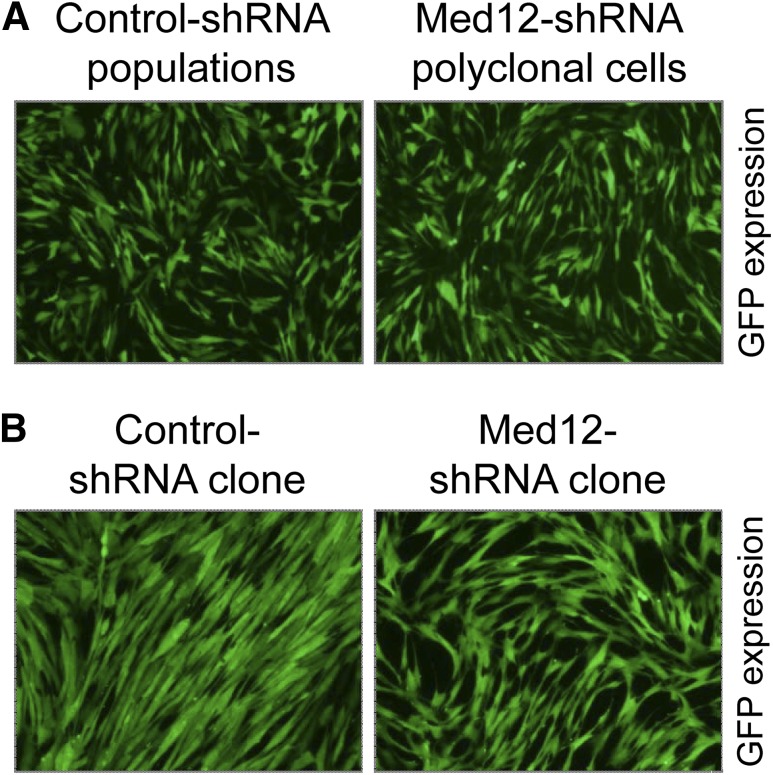
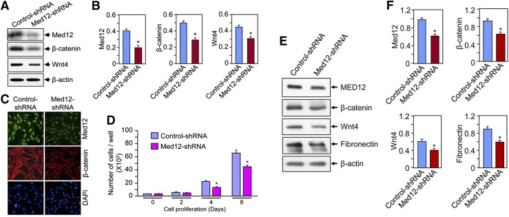
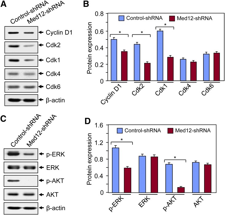
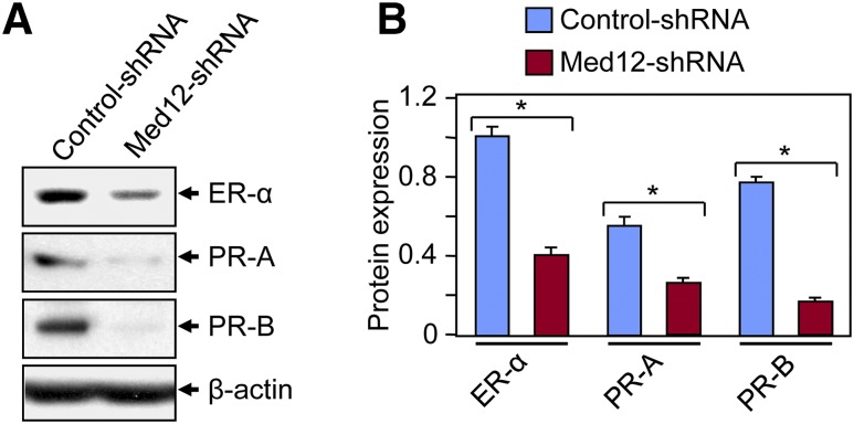
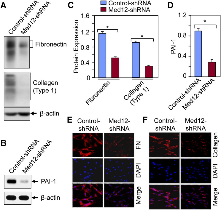
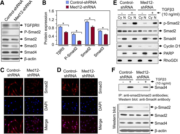
Uterine fibroids, or leiomyoma, are the most common benign tumors in women of reproductive age. In this work, the effect of silencing the mediator complex subunit 12 (Med12) gene in human uterine fibroid cells was evaluated. The role of Med12 in the modulation of Wnt/β-catenin and cell proliferation-associated signaling was evaluated in human uterine fibroid cells. Med12 was silenced in the immortalized human uterine fibroid cell line (HuLM) using a lentivirus-based Med12 gene-specific RNA interference strategy. HuLM cells were infected with lentiviruses carrying Med12-specific short hairpin RNA (shRNA) sequences or a nonfunctional shRNA scrambled control with green fluorescence protein. Stable cells that expressed low levels of Med12 protein were characterized. Wnt/β-catenin signaling, sex steroid receptor signaling, cell cycle-associated, and fibrosis-associated proteins were measured. Med12 knockdown cells showed significantly (P < 0.05) reduced levels of Wnt4 and β-catenin proteins as well as cell proliferation, as compared with scrambled control cells. Med12 knockdown cells also showed reduced levels of cell cycle-associated cyclin D1, Cdk1, and Cdk2 proteins as well as reduced activation of p-extracellular signal-regulated kinase, p-protein kinase B, and transforming growth factor (TGF)-β signaling pathways as compared with scrambled control cells. Moreover, TGF-β-regulated fibrosis-related proteins such as fibronectin, collagen type 1, and plasminogen activator inhibitor-1 were significantly (P < 0.05) reduced in Med12 knockdown cells as compared with scrambled control cells. Together, these results suggest that Med12 plays a key role in the regulation of HuLM cell proliferation through the modulation of Wnt/β-catenin, cell cycle-associated, and fibrosis-associated protein expression.


Figures

Figure 1.
Figure 1.
Generation of Med12 knockdown hUF cells. Med12 gene expression was knocked down in HuLM cells using lentivirus-based RNAi strategy. Lentiviruses harboring Med12 gene–specific short shRNA sequences or nonfunctional scrambled control-shRNA sequence were used to infect HuLM cells and then selected with puromycin (0.5 µg/mL). Stable cell populations expressing higher levels of green fluorescence protein (GFP) are shown (A). These cell populations were further used to isolate stable clones. A stable clone expressing lower levels of Med12 protein (Med12-shRNA) as compared with a control-shRNA clone is shown (B). Photos were taken at magnification of ×100.
Figure 2.
Figure 2.
Effect of Med12 knockdown on Wnt4/β-catenin signaling in hUF cells. (A) Cell lysates prepared from control and Med12 knockdown cells were analyzed by Western blot using anti-Med12, anti-Wnt4, and anti–β-catenin antibodies. β-actin was used as the loading control. (B) The intensity of each protein band was quantified and normalized with corresponding β-actin, as shown. *P < 0.05 as compared with control-shRNA. (C) Immunofluorescence analyses were performed using Med12-shRNA and control-shRNA cells cultured on glass coverslips. Cells were fixed, permeabilized, and stained with monoclonal anti–β-catenin or polyclonal anti-Med12 antibodies, followed by incubation with carbocyanine 3–conjugated anti-mouse secondary or fluorescein isothiocyanate–conjugated anti-rabbit antibodies. Med12 (green) and β-catenin (red) protein expression were monitored by fluorescence microscopy. Cell nuclei were stained with 4′,6-diamidino-2-phenylindole (blue). Photos were taken at magnification of ×200. (D) Cell proliferation assay: Both Med12 knockdown and control-shRNA cells were seeded into 12-well plates and cultured in phenol-free DMEM/F12 medium containing 10% charcoal-stripped fetal bovine serum. Cell proliferation assays were performed by direct cell counting at day 0, day 2, day 4, and day 8. Each data point represents the mean ± SD of triplicate wells (n = 3). *P < 0.05 as compared with the corresponding control-shRNA. (E and F) Protein lysates prepared from scrambled-control and Med12 knockdown fibroid primary cells were analyzed by Western blot using anti-Med12, anti-Wnt4, anti–β-catenin, and anti-fibronectin antibodies. β-actin was used as the loading control. *P < 0.05 as compared with the corresponding control-shRNA.
Figure 3.
Figure 3.
Effect of Med12 knockdown on cell cycle regulatory and growth-promoting pathways in hUF cells. (A) Equal amounts of cell lysates from Med12-shRNA and control-shRNA cells were analyzed by Western blotting using anti-cyclin D1, Cdk1, Cdk2, Cdk4, and Cdk6 antibodies. β-actin Western blot was used as the loading control. (B) The intensity of each protein band was quantified and normalized with corresponding β-actin. *P < 0.05 when compared with control-shRNA. (C) Cell lysates were analyzed by Western blot using antibodies against p-ERK, total ERK, p-AKT, and total AKT. (D) The intensity of each protein band was quantified and normalized with corresponding β-actin. *P < 0.05 when compared with control shRNA.
Figure 4.
Figure 4.
Effect of Med12 knockdown on expression of sex steroid receptors in hUF cells. (A) Protein lysates were prepared from both Med12-shRNA and control-shRNA cells, as described in illustration. Equal amounts of cell lysates were analyzed by Western blotting using anti–ER-α, anti–PR-A, and anti–PR-B antibodies. (B) The intensity of each protein band was quantified and normalized with corresponding β-actin. *P < 0.05 when compared with control shRNA.
Figure 5.
Figure 5.
Effect of Med12 knockdown on expression of fibrosis-associated proteins in hUF cells. Equal amounts of lysates from Med12-shRNA and control-shRNA cells were analyzed by Western blotting using both anti-fibronectin and anti-collagen type 1 (A) and anti–PAI-1 (B) antibodies. β-actin Western blot was used as the loading control. (C and D) The intensity of each protein band was quantified and normalized with corresponding β-actin. *P < 0.05 when compared with control shRNA. (E and F) Immunofluorescence analyses were performed using Med12-shRNA and control-shRNA cells cultured on glass coverslips. Cells were fixed, permeabilized, and incubated with polyclonal anti-fibronectin (E) and anti-collagen type 1 (F) antibodies (1:50 dilution each), followed by incubation with carbocyanine 3–conjugated anti-rabbit secondary antibody. Fibronectin and collagen type 1 expression (red) were monitored by fluorescence microscopy. Cell nuclei were stained with 4′,6-diamidino-2-phenylindole (blue). Photos were taken at magnification of ×200.
Figure 6.
Figure 6.
Effect of Med12 knockdown on TGF-β/Smad signaling in hUF cells. (A) Equal amounts of lysates from Med12-shRNA and control-shRNA cells were analyzed by Western blotting using anti–TGF-βRII, anti–p-Smad2, anti-Smad2, anti-Smad3, and anti-Smad4 antibodies. β-actin Western blot was used as the loading control. (B) The intensity of each protein band was quantified and normalized with corresponding β-actin. *P < 0.05 when compared with control shRNA. (C and D) Immunofluorescence analyses were performed, as described in illustration. Cells were fixed, permeabilized, and incubated with anti-Smad2 and anti-Smad3 (10 µg/mL, each antibody), followed by incubation with carbocyanine 3–conjugated anti-rabbit secondary antibody. Either Smad2 or Smad3 expression (red) was monitored by a fluorescent microscopy. Cell nuclei were stained with 4′,6-diamidino-2-phenylindole (blue). Photos were taken at magnification of ×200. (E) Nuclear (N) and cytoplasmic (Cy) extracts were prepared from both Med12-shRNA and control-shRNA cells treated without or with TGF-β3 (10 ng/mL) for 4 hours. A total of 15 µg each cytoplasmic and nuclear extracts was analyzed by Western blots using anti–p-Smad2, anti-Smad2, anti-Smad4, and anti-cyclin D1 antibodies. Poly(ADP-ribose) polymerase (nuclear) and RhoGDI (cytoplasmic) Western blots were used as to show the purity of the separation. (F) Med12-shRNA and control-shRNA cells were cultured, serum starved, and treated with or without TGF-β3 (10 ng/mL) for 4 hours. Equal amounts of cell lysates (1 mg) were used for the immunoprecipitation (IP) assay with both anti-Smad2 and anti-Smad3 antibodies (2 µg each) for 2.5 hours at 4°C. A total of 25 µL protein G–Sepharose beads was added and then incubated for another hour. Immune complex was washed 4 times (5 minutes each), and thereafter Western blot analyses were performed using anti-Smad4 antibody (top panel). Cell lysates were verified by Western blots for expression of p-Smad2, Smad2, and Smad4 proteins. β-actin Western blot was used as the loading control (bottom panels).

Similar articles

Cited by

References

    1. Walker CL, Stewart EA. Uterine fibroids: the elephant in the room. Science. 2005;308(5728):1589–1592. - PubMed
    1. Marsh EE, Bulun SE. Steroid hormones and leiomyomas. Obstet Gynecol Clin North Am. 2006;33(1):59–67. - PubMed
    1. Payson M, Leppert P, Segars J. Epidemiology of myomas. Obstet Gynecol Clin North Am. 2006;33(1):1–11. - PMC - PubMed
    1. Laughlin SK, Schroeder JC, Baird DD. New directions in the epidemiology of uterine fibroids. Semin Reprod Med. 2010;28(3):204–217. - PMC - PubMed
    1. Gupta S, Jose J, Manyonda I. Clinical presentation of fibroids. Best Pract Res Clin Obstet Gynaecol. 2008;22(4):615–626. - PubMed

MeSH terms