Radiopacity of alloplastic bone grafts measured with cone beam computed tomography: An analysis in rabbit calvaria
- PMID: 27968706
- PMCID: PMC5341780
- DOI: 10.17305/bjbms.2016.1482
Radiopacity of alloplastic bone grafts measured with cone beam computed tomography: An analysis in rabbit calvaria
Abstract
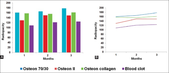
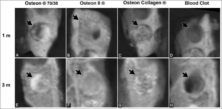
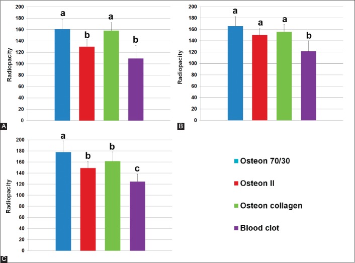
Availability of adequate bone structure for dental implants is still a problem in dentistry. Alloplastic grafts, which promote bone regeneration, are used as bone substitutes in orthopedic and oral surgical procedures. The aim of this study was to evaluate the radiopacity of three different synthetic bone grafts in rabbit calvaria, over 3 months, using cone beam computed tomography (CBCT). Four critical-size defects were made on the calvaria of 11 rabbits. The lesions were classified into three groups according to the alloplastic grafts they received: Osteon® 70/30, Osteon collagen®, and Osteon II® groups. The fourth group received blood clot, and served as a control. The bone samples were collected and analyzed with CBCT after the 1st, 2nd, and 3rd month. One month after surgery, the lesions that received Osteon® 70/30 and Osteon collagen® grafts showed the highest radiopacity compared to the lesions with Osteon II® and blood clot. After the 2nd month, the radiopacity values between the three groups that received the grafts were more similar compared to the group with blood clot. After the 3rd month, the lesions with Osteon® 70/30 graft showed the highest radiopacity values, followed by Osteon collagen® and Osteon II® groups. The group that received blood clot showed the lowest radiopacity values. In conclusion, the grafts used in this study had higher radiopacity values compared to blood clot. Among the grafts used, the Osteon® 70/30 graft showed the highest radiopacity values in the 3-month period.
Figures
References
-
- Ogose A, Hotta T, Kawashima H, Kondo N, Gu W, Kamura T, et al. Comparison of hydroxyapatite and beta tricalcium phosphate as bone substitutes after excision of bone tumors. J Biomed Mater Res B Appl Biomater. 2005;72(1):94–101. http://dx.doi.org/10.1002/jbm.b.30136. - PubMed
-
- Denry I, Kuhn LT. Design and characterization of calcium phosphate ceramic scaffolds for bone tissue engineering. Dent Mater. 2016;32(1):43–53. http://dx.doi.org/10.1016/j.dental.2015.09.008. - PMC - PubMed
-
- Larsson S. Calcium phosphates: What is the evidence? J Orthop Trauma. 2010;24(Suppl 1):S41–5. DOI: 10.1097/BOT.0b013e3181cec472. - PubMed
-
- Yamada S, Heymann D, Bouler JM, Daculsi G. Osteoclastic resorption of calcium phosphate ceramics with different hydroxyapatite/beta-tricalcium phosphate ratios. Biomaterials. 1997;18(15):1037–41. http://dx.doi.org/10.1016/S0142-9612(97)00036-7. - PubMed
MeSH terms
Substances
LinkOut - more resources
Full Text Sources
Other Literature Sources
Medical