

Effects of oncostatin M on cell proliferation and osteogenic differentiation in C3H10T1/2
- PMID: 27973390
- PMCID: PMC5259579
Effects of oncostatin M on cell proliferation and osteogenic differentiation in C3H10T1/2
Abstract
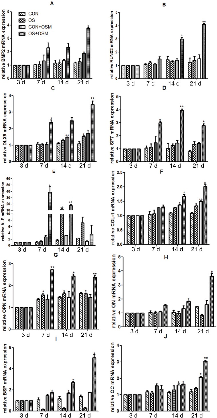
Objective: To explore the effects of protein factor Oncostatin M (OSM), a member of the Interleukin-6 (IL-6) family on cell proliferation, osteogenic differentiation and mineralization.
Materials and methods: Basal nutrient solutions of different concentrations of OSM (0, 5, 10, 20, 40, 80 ng/ml) were used. In order to divide embryonic origin between mesenchymal stem cells C3H10T1/2 of in vitro cultured mice, and the effects of in vitro proliferation efficiencies of C3H10T1/2 cells of different concentrations of OSM, the C3H10T1/2 cells were divided into four groups: (1) Basal nutrient solution group (negative control); (2) Osteogenesis induced liquid group (positive control); (3) OSM (20 ng/ml) group; (4) Experimental group (osteogenesis induced liquid + OSM (20 ng/ml)). The expressions levels of relevant osteogenesis and mineralization genes were detected.
Results: OSM had several effects on promoting the proliferation of embryonic origin mesenchymal stem cells C3H10T1/2 with respect to time of exposure as well as concentrations. In the present study, it has been shown that when the concentration of OSM is 20 ng/ml, the effects of promoting proliferation are most obvious. OSM can induce osteogenic differentiation of C3H10T1/2, make the process of osteogenic differentiation in advance, and promote the formation of end-stage calcium deposits and mineralized nodule, and osteogenic differentiation of C3H10T1/2 is finally achieved.
Conclusion: OSM can promote the proliferation of C3H10T1/2, and induce its osteogenic differentiation and end-stage mineralization.
Conflict of interest statement
The authors have no conflict of interest.
Figures


References
-
- Case ND, Duty AO, Ratcliffe A, Muller R, Guldberg RE. Bone formation on tissue-engineered cartilage constructs in vivo: Effects of chondrocyte viability and mechanical loading. Tissue Eng. 2003;9:587–596. - PubMed
-
- Bruder SP, Kraus KH, Goldberg VM, Kadiyala S. The effect of implants loaded with autologous mesenchymal stem cells on the healing of canine segmental bone defects. J Bone Joint Surg Am. 1998;80:985–996. - PubMed
-
- Giannoudis PV, Hak D, Sanders D, Donohoe E, Tosounidis T, Bahney C. Inflammation, bone healing, and Anti-Inflammatory drugs: An update. J Orthop Trauma. 2015;29 Suppl(12):S6–S9. - PubMed
-
- Moxham JP. Oncostatin-M enhances osteoinduction in a rabbit critical calvarial defect model. Laryngoscope. 2007;117:1790–1797. - PubMed
MeSH terms
Substances
LinkOut - more resources
Full Text Sources
Other Literature Sources