. 2016 Dec 10;8(12):112.
doi: 10.3390/cancers8120112.

Comparative miRNA Analysis of Urine Extracellular Vesicles Isolated through Five Different Methods

Affiliations

Comparative miRNA Analysis of Urine Extracellular Vesicles Isolated through Five Different Methods

Felix Royo et al. Cancers (Basel). .

Abstract

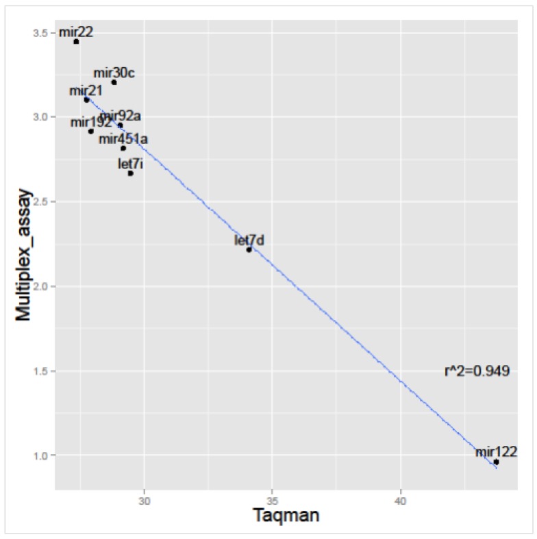
Urine extracellular vesicles are a valuable low-invasive source of information, especially for the cells of the genitourinary tract. In the search for biomarkers, different techniques have been developed to isolate and characterize the cargo of these vesicles. In the present work, we compare five of these different isolation methods (three commercial isolation kits, ultracentrifugation, and lectin-based purification) and perform miRNA profiling using a multiplex miRNA assay. The results showed high correlation through all isolation techniques, and 48 out of 68 miRNAs were detected above the detection limit at least 10 times. The results obtained by multiplex assay were validated through Taqman qPCR. In addition, using this technique combined with a clinically friendly extracellular vesicle (uEV)-enrichment method, we performed the analysis of selected miRNAs in urine from patients affected with bladder cancer, benign prostate hyperplasia, or prostate cancer. Importantly, we found that those miRNAs could be detected in almost 100% of the samples, and no significant differences were observed between groups. Our results support the feasibility of analyzing exosomes-associated miRNAs using a methodology that requires a small volume of urine and is compatible with a clinical environment and high-throughput analysis.

Keywords: exosomes; extracellular vesicles; isolation methods; miRNA; urine.


Conflict of interest statement

The authors declare no conflict of interest.

Figures

Figure 1
Figure 1
Heatmap analysis of multiplex miRNA assay. The color code corresponds to the signal intensity obtained in the assay, and samples were grouped by isolation method. Each square corresponds to a single miRNA and sample. CEN: ultracentrifugation; EXQ: Exoquick-TC; INV: Total Exosome Isolation Solution; LEC: lectin-based purification; NOR: Urine Exosome RNA Isolation Kit.
Figure 2
Figure 2
Correlation matrix between different methods. The r coefficients show that LEC purification performed slightly different than the other methods. All the correlations are highly significant (p < 0.001).
Figure 3
Figure 3
ANOVA analysis of the performance of the different methods for (a) hsa-mir-30c-5p and (b) hsa-mir-92a-3p detection. Note: in panel (a), NOR is significantly different from the other treatments, while in panel (b) INV is different from the rest of the treatments (p < 0.05, by post-hoc comparisons using Bonferroni correction).
Figure 4
Figure 4
Percentage of detection for each miRNA, employing (a) Taqman qPCR or (b) Multiplex Circulating miRNA Assay (Abcam).
Figure 4
Figure 4
Percentage of detection for each miRNA, employing (a) Taqman qPCR or (b) Multiplex Circulating miRNA Assay (Abcam).
Figure 5
Figure 5
Correlation between signal intensity (Log10 transformed) achieved by multiplex miRNA assay and Ct values obtained by Taqman qPCR, using the same RNAs.
Figure 6
Figure 6
miRNAs analysis of extracellular vesicles (uEVs)-enriched preparations obtained of different genitourinary track pathologies. NT (non-tumoral, patients without malignancies), BCa (bladder cancer), PCa (prostate cancer), and BPH (benign prostate hyperplasia). Fold changes were calculated using cel-mir-39c-3p as housekeeping, and the data was normalized against the average of the NT group, so the average of the group is 1 for each miRNA. ANOVA did not find any significant differences amongst groups.

References

    1. Pisitkun T., Shen R.F., Knepper M.A. Identification and proteomic profiling of exosomes in human urine. Proc. Natl. Acad. Sci. USA. 2004;101:13368–13373. doi: 10.1073/pnas.0403453101. - DOI - PMC - PubMed
    1. Hoorn E.J., Pisitkun T., Zietse R., Gross P., Frokiaer J., Wang N.S., Gonzales P.A., Star R.A., Knepper M.A. Prospects for urinary proteomics: Exosomes as a source of urinary biomarkers. Nephrology. 2005;10:283–290. doi: 10.1111/j.1440-1797.2005.00387.x. - DOI - PubMed
    1. Miranda K.C., Bond D.T., McKee M., Skog J., Paunescu T.G., da Silva N., Brown D., Russo L.M. Nucleic acids within urinary exosomes/microvesicles are potential biomarkers for renal disease. Kidney Int. 2010;78:191–199. doi: 10.1038/ki.2010.106. - DOI - PMC - PubMed
    1. Cheng Y., Wang X., Yang J., Duan X., Yao Y., Shi X., Chen Z., Fan Z., Liu X., Qin S., et al. A translational study of urine miRNAs in acute myocardial infarction. J. Mol. Cell. Cardiol. 2012;53:668–676. doi: 10.1016/j.yjmcc.2012.08.010. - DOI - PMC - PubMed
    1. Chen C.L., Lai Y.F., Tang P., Chien K.Y., Yu J.S., Tsai C.H., Chen H.W., Wu C.C., Chung T., Hsu C.W., et al. Comparative and targeted proteomic analyses of urinary microparticles from bladder cancer and hernia patients. J. Proteome Res. 2012;11:5611–5629. doi: 10.1021/pr3008732. - DOI - PubMed