. 2016 Dec 14;11(12):e0168269.
doi: 10.1371/journal.pone.0168269. eCollection 2016.

The In Vitro Effect of Acidic-Pepsin on Nuclear Factor KappaB Activation and Its Related Oncogenic Effect on Normal Human Hypopharyngeal Cells

Affiliations

The In Vitro Effect of Acidic-Pepsin on Nuclear Factor KappaB Activation and Its Related Oncogenic Effect on Normal Human Hypopharyngeal Cells

Clarence T Sasaki et al. PLoS One. .

Abstract

Background: Extra-esophageal carcinogenesis has been widely discussed in relation to the chronic effects of laryngopharyngeal reflux and most prominently with pepsin historically central to this discussion. With refluxate known to include gastric (pepsin) and duodenal (bile) fluids, we recently demonstrated the mechanistic role of NF-κB in mediating the preneoplastic effects of acidic-bile. However, the role of pepsin in promoting hypopharyngeal premalignant events remains historically unclear. Here, we investigate the in vitro effect of acidic-pepsin on the NF-κB oncogenic pathway to better define its potential role in hypopharyngeal neoplasia.

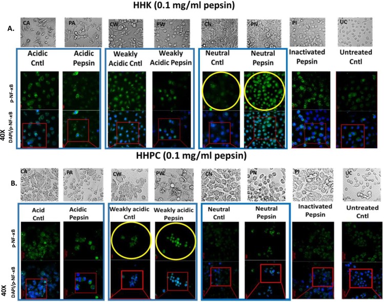
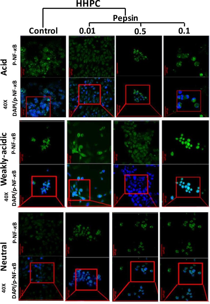
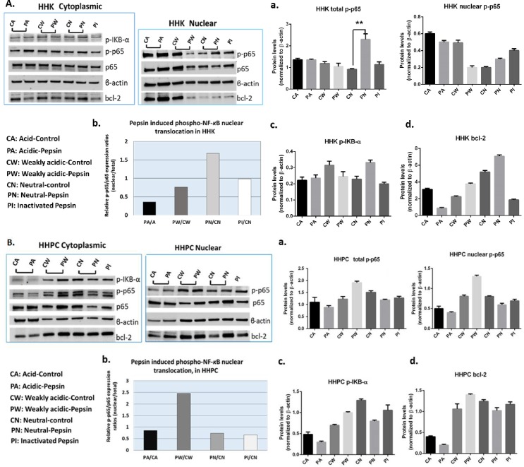
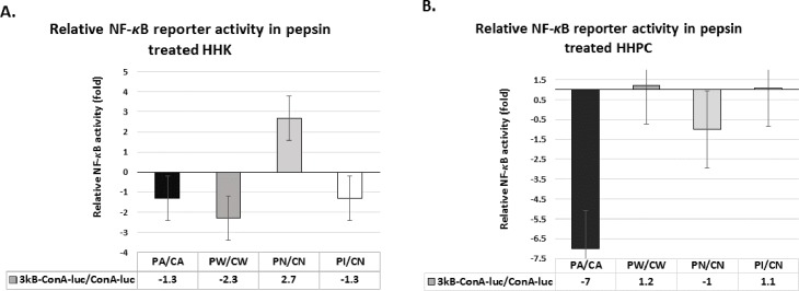
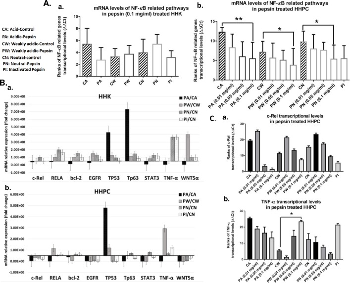
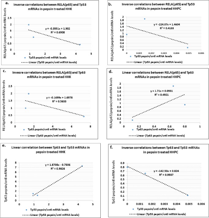
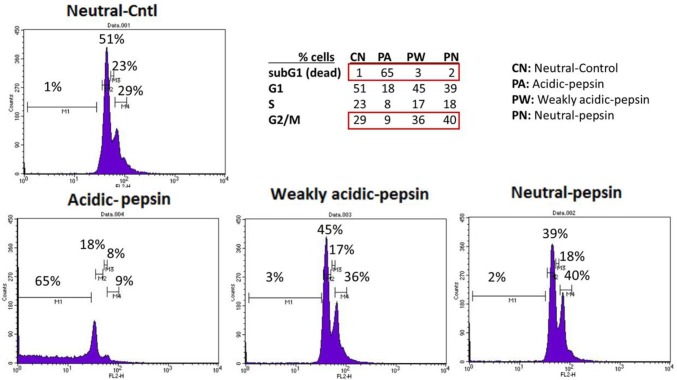
Methods: Human hypopharyngeal primary cells (HHPC) and keratinocytes (HHK) were repetitively exposed to physiologic pepsin concentrations (0.1 mg/ml) at pH 4.0, 5.0 and 7.0. Cellular localization of phospho-NF-κB and bcl-2 was determined using immunofluorescence and western blotting. NF-κB transcriptional activity was tested by luc reporter and qPCR. Analysis of DNA content of pepsin treated HHK and HHPC was performed using Fluorescence-activated-cell sorting assay. To explore a possible dose related effect, pepsin concentration was reduced from 0.1 to 0.05 and 0.01 mg/ml.

Results: At physiologic concentration, acidic-pepsin (0.1 mg/ml at pH 4.0) is lethal to most normal hypopharyngeal cells. However, in surviving cells, no NF-κB transcriptional activity is noted. Acidic-pepsin fails to activate the NF-κB or bcl-2, TNF-α, EGFR, STAT3, and wnt5α but increases the Tp53 mRNAs, in both HHPC and HHK. Weakly acidic-pepsin (pH 5.0) and neutral-pepsin (pH 7.0) induce mild activation of NF-κB with increase in TNF-α mRNAs, without oncogenic transcriptional activity. Lower concentrations of pepsin at varying pH do not produce NF-κB activity or transcriptional activation of the analyzed genes.

Conclusion: Our findings in vitro do not support the role of acidic-pepsin in NF-κB related hypopharyngeal carcinogenesis.


Conflict of interest statement

The authors have declared that no competing interests exist.

Figures

Fig 1
Fig 1. Effect of pepsin in p-NF-κB nuclear localization, in human hypopharyngeal keratinocytes (HHK) and human hypopharyngeal primary cells (HHPC).
Immunofluorescence (IF) staining of phospho-NF-κB (p-p65 S529) is demonstrated in (A) HHK and (B) HHPC treated by 0.1 mg/ml pepsin at different pH (acid, pH 4.0; weakly acidic, pH 5.0 and neutral, pH 7.0) and corresponding controls. Acidic-pepsin treated HHK and HHPC demonstrates mainly cytoplasmic localization of p-NF-κB. In contrast, neutral-pepsin treated HHK and weakly acidic-pepsin treated HHPC results in an intense nuclear localization of p-NF-κB.
Fig 2
Fig 2. Varying pepsin concentrations at different pH do not affect the nuclear localization of p-NF-κB, in human hypopharyngeal primary cells (HHPC).
Immunofluorescence (IF) staining of phospho-NF-κB (p-p65 S529) is demonstrated in HHPC treated by 0.01, 0.5 and 01 mg/ml of pepsin at different pH (acid, pH 4.0; weakly acidic, pH 5.0 and neutral, pH 7.0) and corresponding controls.
Fig 3
Fig 3. Effect of pepsin in NF-κB activation and bcl-2 overexpression, in human hypopharyngeal keratinocytes (HHK) and human hypopharyngeal primary cells (HHPC).
Western blot analysis for NF-κB (p65), p-NF-κB (p-p65 S529), p-IKB-α (Ser32/36) and bcl-2 protein levels demonstrates that acidic-pepsin does not induce NF-κB activation or bcl-2 overexpression in treated (A) HHK and (B) HHPC. Columns of the graph correspond to (a) total and nuclear p-NF-κB (p-p65) levels, (b) pepsin-induced nuclear p-NF-κB (p65 S529) translocation values (p-p65/p65 nuclear/total ratios), (c) cytoplasmic p-IKB-α and (d) cytoplasmic/nuclear bcl-2 protein levels (ONE-WAY ANOVA; Kruskal-Wallis; GraphPad Prism 6.0). Mean ±SD of three independent experiments.
Fig 4
Fig 4
Luciferase assay demonstrates the effect of pepsin in NF-κB transcriptional activity in (A) HHK and (B) HHPC. Acidic-pepsin (pH 4.0) does not induce NF-κB transcriptional activity in treated human normal hypopharyngeal cells. Columns represent NF-κB relative transcriptional activity (NF-κB responsive luciferase reporter/control luciferase reporter). 3kB-ConA-luc: NF-κB responsive luciferase reporter; ConA-luc: control luciferase reporter; 3kB-ConA-luc/ConA-luc: NF-κB responsive luciferase reporter/control luciferase reporter. CA: Acid-Control; PA: Acidic-Pepsin; CW: Weakly Acidic-Control; PW: Weakly Acidic-Pepsin; CN: Neutral-Control; PN: Neutral-Pepsin; PI: Inactivated-Pepsin.
Fig 5
Fig 5. Quantitative real-time PCR demonstrates the effect of pepsin in transcriptional activation of NF-κB and related oncogenic pathway, in treated human hypopharyngeal keratinocytes (HHK) and human hypopharyngeal primary cells (HHPC).
A. Transcriptional levels of all the analyzed NF-κB related genes (normalized to hGAPDH) are demonstrated in pepsin and control treated (a) HHK and (b) HHPC. Transcriptional levels of all the analyzed genes were significantly lower in HHPC exposed to 0.1 mg/ml of pepsin, compared to their corresponding controls. (ONE-WAY ANOVA, by Freidman; Dunn’s multiple comparisons test; *p<0.01; **p<0.001). B. Columns correspond to relative (pepsin/control) normalized transcriptional levels of each bcl-2, EGFR, c-REL, RELA(p65), Tp63, STAT3, wnt5α, TNF-α, and Tp53 gene, in treated (a) HHK and (b) HHPC. C. Columns correspond to transcriptional levels of (a) c-REL and (b) TNF-α in HHPC exposed to varying pepsin-concentrations and pH, and corresponding controls. (ONE-WAY ANOVA, Kruskal-Wallis; Dunn’s multiple comparison test; GraphPad Prism 6 software). (CA: Acid-Control; PA: Acidic-Pepsin; CW: Weakly Acidic-Control; PW: Weakly Acidic-Pepsin; CN: Neutral-Control; PN: Neutral-Pepsin; PI: Inactivated-Pepsin).
Fig 6
Fig 6. Diagrams show significant correlations by Pearson of relative (pepsin/cntl) expression between NF-κB and related genes in treated HHK and HHPC.
Correlation is demonstrated between (a) RELA(p65) and Tp53 mRNAs in HHK, (b) RELA(p65) and Tp53 mRNAs in HHPC, (c) RELA(p65) and Tp63 mRNAs HHK, (d) RELA(p65) and Tp63 mRNAs in HHPC, as well as between (e) Tp53 and Tp63 mRNAs in HHK and (f) Tp53 and Tp63 mRNAs in HHPC (by Pearson; p-value<0.05).
Fig 7
Fig 7. Fluorescence-activated cell sorting assay (FACS) demonstrates pronounced cell death (subG1 population) in acidic-pepsin treated human hypopharyngeal keratinocytes (HHK).
FACS shows that less acidic pH (≥5.0) is less toxic to pepsin-treated HHK, resulting in higher percentage (%) of cells in G2/M phase relative to acidic-pepsin (pH 4.0).

References

    1. American Cancer Society. Cancer reference information. Available from URL: http://m.cancer.org/cancer/laryngealandhypopharyngealcancer/detailedguid... [accessed February, 2016].
    1. American Cancer Society. Cancer reference information. Available from URL: http://m.cancer.org/cancer/laryngealandhypopharyngealcancer/detailedguid... [acessed February, 2016].
    1. Jégu J, Binder-Foucard F, Borel C, Velten M. Trends over three decades of the risk of second primary cancer among patients with head and neck cancer. Oral Oncol. 2013; 49(1):9–14. 10.1016/j.oraloncology.2012.06.018 - DOI - PubMed
    1. Hashibe M, Boffetta P, Zaridze D, Shangina O, Szeszenia-Dabrowska N, Mates D, et al. Contribution of tobacco and alcohol to the high rates of squamous cell carcinoma of the supraglottis and glottis in Central Europe. Am J Epidemiol. 2007; 165(7):814–820. 10.1093/aje/kwk066 - DOI - PubMed
    1. McQuaid KR, Laine L, Fennerty MB, Souza R, Spechler SJ. Systematic review: the role of bile acids in the pathogenesis of gastro-oesophageal reflux disease and related neoplasia. Aliment Pharmacol Ther. 2011; 34:146–165. 10.1111/j.1365-2036.2011.04709.x - DOI - PubMed

MeSH terms