Review
. 2017 Jun;284(11):1590-1605.
doi: 10.1111/febs.13990. Epub 2017 Jan 6.

New insights into the evolutionary origins of the recombination-activating gene proteins and V(D)J recombination

Affiliations
Review

New insights into the evolutionary origins of the recombination-activating gene proteins and V(D)J recombination

Lina Marcela Carmona et al. FEBS J. 2017 Jun.

Abstract

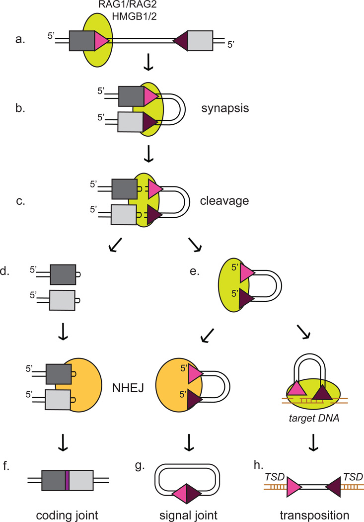
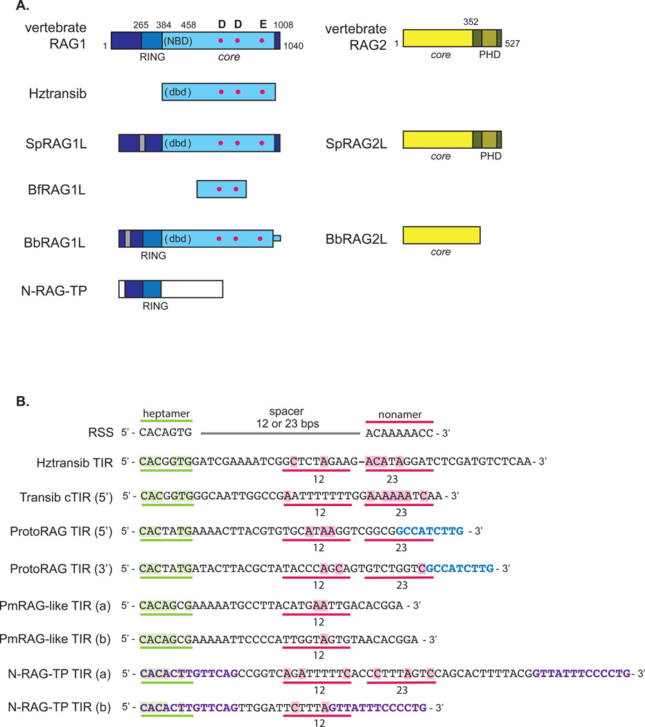
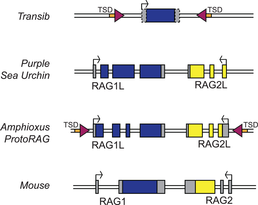
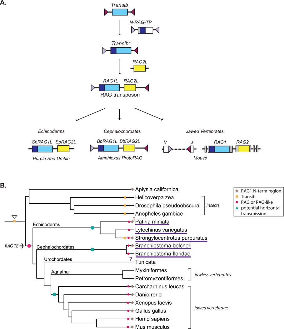
The adaptive immune system of jawed vertebrates relies on V(D)J recombination as one of the main processes to generate the diverse array of receptors necessary for the recognition of a wide range of pathogens. The DNA cleavage reaction necessary for the assembly of the antigen receptor genes from an array of potential gene segments is mediated by the recombination-activating gene proteins RAG1 and RAG2. The RAG proteins have been proposed to originate from a transposable element (TE) as they share mechanistic and structural similarities with several families of transposases and are themselves capable of mediating transposition. A number of RAG-like proteins and TEs with sequence similarity to RAG1 and RAG2 have been identified, but only recently has their function begun to be characterized, revealing mechanistic links to the vertebrate RAGs. Of particular significance is the discovery of ProtoRAG, a transposon superfamily found in the genome of the basal chordate amphioxus. ProtoRAG has many of the sequence and mechanistic features predicted for the ancestral RAG transposon and is likely to be an evolutionary relative of RAG1 and RAG2. In addition, early observations suggesting that RAG1 is able to mediate V(D)J recombination in the absence of RAG2 have been confirmed, implying independent evolutionary origins for the two RAG genes. Here, recent progress in identifying and characterizing RAG-like proteins and the TEs that encode them is summarized and a refined model for the evolution of V(D)J recombination and the RAG proteins is presented.

Keywords: ProtoRAG; RAG-like proteins; RAG1; RAG2; Transib; V(D)J recombination; evolution; transposition.


Figures

Figure 1
Figure 1. Outline of RAG DNA cleavage reaction and products
RAG mediated cleavage begins with the binding of RAG1/RAG2 along with HMGB1/2 (green oval) to a single RSS (triangle) (a). Synapsis with a partner RSS (b) allows for the cleavage reaction to proceed (c), specifically enabling the hairpin formation step of the nick-hairpin cleavage mechanism. Note that the reaction is most efficient with one 12RSS (pink triangle) and one 23RSS (purple triangle), a restriction known as the 12/23 rule. After cleavage, two types of products are generated. The hairpins are present on the DNA ends containing the gene segments, known as the coding ends (d). They are opened and processed by the NHEJ pathway (orange oval) to generate the coding joint (f). The blunt DNA ends containing the RSSs, known as the signal ends (e), are also usually handed over to the NHEJ pathway to be ligated together, forming the signal joint (g). See [3] for a more detailed summary of this reaction. However, the signal ends also have the potential to undergo a transposition reaction in which the free 3’ hydroxyl groups attack a target piece of DNA (brown) with a stagger of 5 base pairs between the top and bottom strands, generating the TSD and integrating the RSSs and intervening DNA into the target DNA (h). This transposition reaction can lead to genome instability and is very rare in vivo.
Figure 2
Figure 2. Comparison of RAG and RAG-like proteins: domain structure and DNA binding sequences
A. The vertebrate RAG1 protein can be truncated to a catalytically active core region (amino acids 384 to 1008 in the mouse protein) (light blue). This contains DNA binding regions, such as the nonamer binding domain (NBD), as well as the three residues that make up the catalytic triad (D600, D708, and E962 in the mouse protein) (red dots). The N-terminal noncore region of RAG1 contains a RING/Zinc finger (RING) that coordinates four zinc atoms [26, 27]. Transib contains sequence similarity only to the core of RAG1, and while it may contain a DNA binding domain (dbd), it but does not have strong sequence similarity to the RAG1 NBD [48]. BfRAG1L contains sequence similarity to only a central portion of RAG1 core, and is therefore missing the last residue of the catalytic triad [54]. BbRAG1L contains sequence similarity that extends into the N-terminal noncore region of RAG1, spanning almost all of the RING/Zinc finger region, but similarity is higher in the core [57], while SpRAG1L contains only limited sequence similarity to the RAG1 RING/Zinc finger region [51]. These two proteins might contain a dbd positioned similarly to the NBD, but do not have sequence similarity to the RAG1 NBD. While HzTransib transposase also lacks sequence similarity to the NBD, some Transib transposases contain NBD-like sequences [53]. Both SpRAG1L and BbRAG1L also contain a series of repeats (gray) but in different locations in their N-terminal regions. Note that the core region of BbRAG1L extends to the C-terminus of the protein, unlike vertebrate RAG1. N-RAG-TP contains sequence similarity solely to the RAG1 N-terminal noncore region. The vertebrate RAG2 protein can be truncated to the core region (amino acids 1 to 352 in the mouse protein) (light yellow), consisting of six kelch repeats which fold into a six-bladed beta propeller, and the noncore PHD (dark yellow). Purple sea urchin and amphioxus contain RAG2-like proteins, but BbRAG2L is missing the PHD finger. The RAG-like proteins from the green sea urchin and bat starfish are not included as their sequences are not well established [53]. No RAG2-like protein has been reported in Aplysia. B. The RSS consists of a well conserved heptamer (green), a spacer that is either 12 or 23 base pairs, and a well conserved nonamer (pink). The only invariant residues in the RSS are the first three (5’-CAC) of the heptamer. The consensus sequence of the Hztransib TIRs [49], the Transib family TIRs [48], and the 5’ and 3’ TIRs reported for ProtoRAG [57], the bat star (PmRAG-like) [53], and N-RAG-TP from Aplysia are aligned. The corresponding location of the heptamer is underlined in green, and the corresponding location of the nonamer given either a 12 or 23 base pair spacer is underlined in pink. ProtoRAG TIRs contain a 9 base pair sequence (blue) that is well conserved and is separated by either 27 or 31 base pairs from the heptamer while N-RAG-TP has two conserved, distinct tridecamer sequences (purple) separated by either 39 or 13 base pairs. Note that all TIRs of RAG-like TEs have strong conservation of residues in the heptamer (residues highlighted in green), and that alignment of the Hztransib TIR has been shifted by 1 base pair for better alignment of the nonamer. Residues with identity to the nonamer are highlighted in pink.
Figure 3
Figure 3. RAG and RAG-like genes
The structure of the vertebrate RAG locus and of several RAG-like genes or TEs is depicted schematically [51, 57]. Transib and the ProtoRAG element from amphioxus contain TIRs (purple triangles) and 5 base pair TSDs (orange boxes) [49, 57]. Transib shares sequence similarity to RAG1 and does not contain a RAG2-like gene [48]. The coding regions for RAG1/RAG1-like genes and RAG2/RAG2-like genes are in blue and yellow respectively, while gray boxes represent untranslated portions of exons. Start sites of transcription are represented with arrows. The 5’ and 3’ untranslated regions of the Transib transposase gene are not well defined (hatch-marked boxes).
Figure 4
Figure 4. Model for the evolution of the RAG proteins and transmission of the ancestral RAG transposon
A. We propose that RAG1 originated from an ancient relative of the Transib TEs. A recombination event between this ancient element and a relative of N-RAG-TP could have generated a new element, Transib*, containing an open reading frame containing both the noncore N-terminal domain and core region of the RAG1 precursor, as well as one new TIR. Subsequent acquisition of a RAG2-like gene by Transib* led to the emergence of the RAG transposon that gave rise to the RAG and RAG-like genes found in echinoderms, cephalochordates, and jawed vertebrates. In the sea urchin, the TIRs of the RAG-like element appear to have been lost while in amphioxus, the TIRs have been maintained and the ProtoRAG element is active in vitro and may retain activity in vivo. In jawed vertebrates, the TIRs went on to become the RSSs after being inserted into a gene (the ancestral split receptor gene) that gave rise into the antigen receptor loci as predicted by the transposon/split receptor gene hypothesis [38, 42, 43]. RAG1/RAG1-like and RAG2/RAG2-like genes are in blue and yellow respectively, and the N-terminal noncore region of RAG1 is in dark blue. TIRs and RSSs are purple triangles. No attempt has been made to depict the origins of RAG2 C-terminal region containing the PHD, although the most parisomonious hypothesis is that it was present in RAG2L of the RAG transposon. B. We propose that the RAG transposon emerged in the genome of a common deuterostome ancestor (arrow and large pink dot), leading to the existence of RAG-like genes in numerous lineages (small pink dots). The widespread presence of these genes can be accounted for through vertical transmission of the RAG TE and loss of RAG-like genes in the jawless fish and at least some tunicates. A model involving horizontal transmission is also possible, and in this case, three independent integration events (teal dots) of a RAG TE would have been required to explain the presence of RAG-like genes in various lineages. Orange squares mark the presence of Transib TEs [48] and the presumed presence of ancient Transib TEs (open arrow head) prior to the divergence of protostomes and deuterostomes. Organisms in which RAG1-like genes have been reported are underlined in purple. The presence or absence of RAG1-like genes in tunicates has not been definitively determined (purple question mark). The N-terminal noncore domain may have originated from a relative of the N-RAG-TP element reported in Aplysia californica. Species with a RAG1-like protein containing the N-terminal domain are marked with a brown diamond. It is not clear whether the N-terminal region of the bat star RAG1-like has homology to RAG1 (brown question mark).

Similar articles

Cited by

References

    1. Schatz DG, Oettinger MA, Baltimore D. The V(D)J recombination activating gene, RAG-1. Cell. 1989;59:1035–1048. - PubMed
    1. Oettinger MA, Schatz DG, Gorka C, Baltimore D. RAG-1 and RAG-2, adjacent genes that synergistically activate V(D)J recombination. Science. 1990;248:1517–1523. - PubMed
    1. Schatz DG, Swanson PC. V(D)J recombination: mechanisms of initiation. Annu Rev Genet. 2011;45:167–202. - PubMed
    1. Kim MS, Lapkouski M, Yang W, Gellert M. Crystal structure of the V(D)J recombinase RAG1-RAG2. Nature. 2015;518:507–511. - PMC - PubMed
    1. Ru H, Chambers MG, Fu TM, Tong AB, Liao M, Wu H. Molecular Mechanism of V(D)J Recombination from Synaptic RAG1-RAG2 Complex Structures. Cell. 2015;163:1138–1152. - PMC - PubMed

Publication types

MeSH terms