. 2016 Dec 13;17(11):2966-2978.
doi: 10.1016/j.celrep.2016.11.050.

Downregulation of a GPCR by β-Arrestin2-Mediated Switch from an Endosomal to a TGN Recycling Pathway

Affiliations

Downregulation of a GPCR by β-Arrestin2-Mediated Switch from an Endosomal to a TGN Recycling Pathway

Nazish Abdullah et al. Cell Rep. .

Abstract

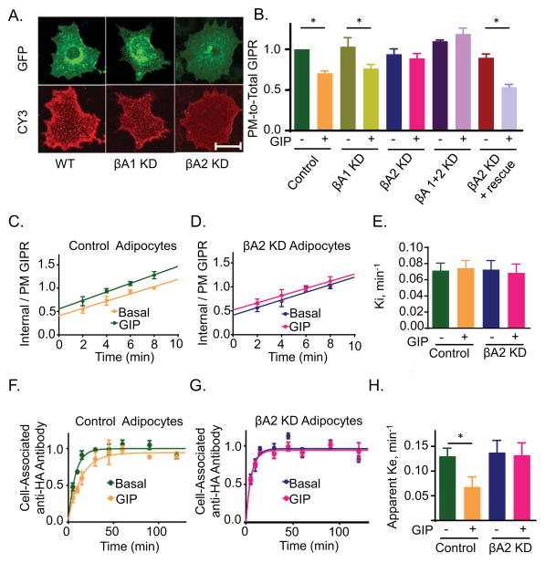
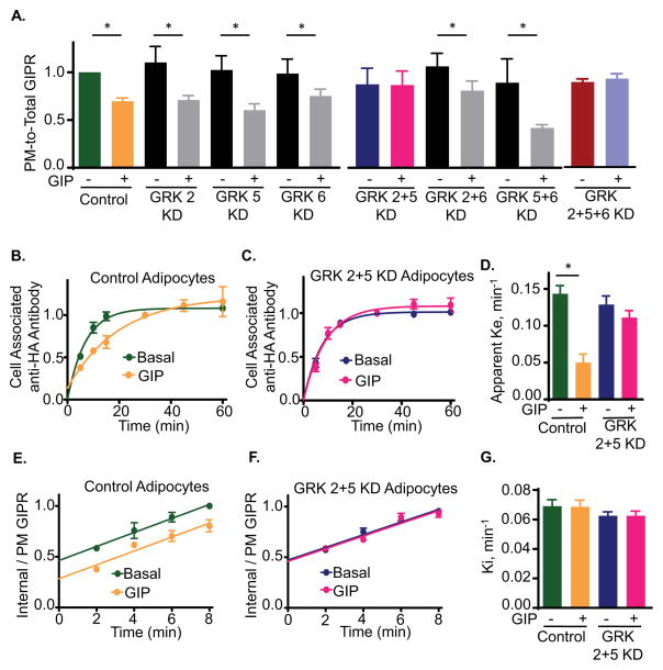
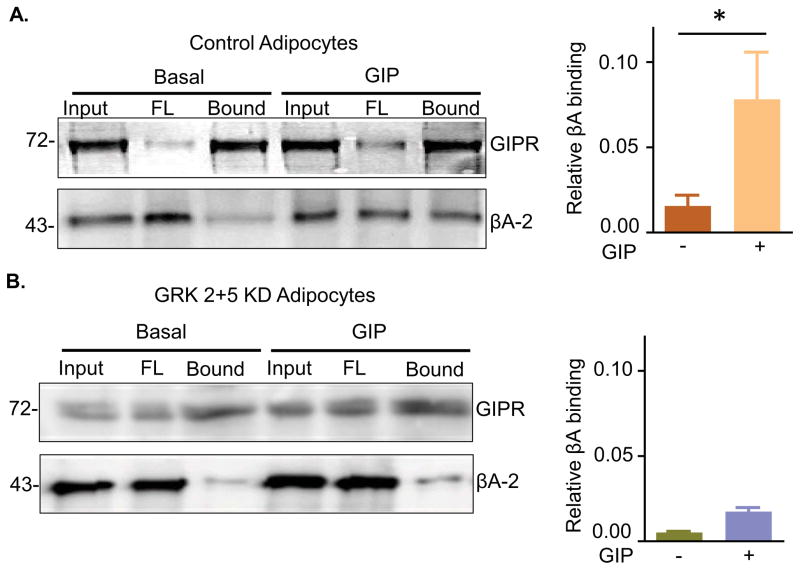
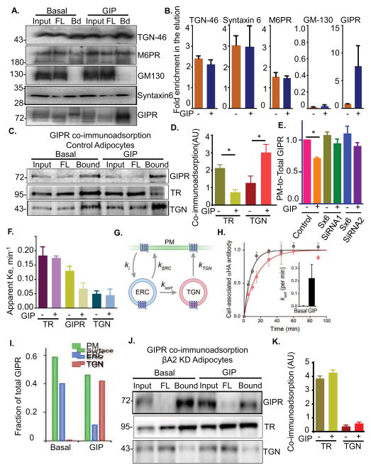
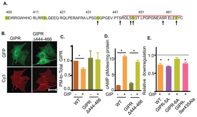
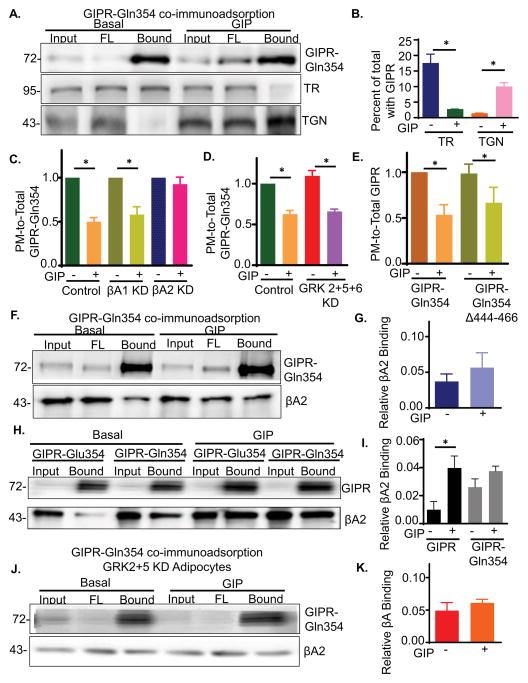
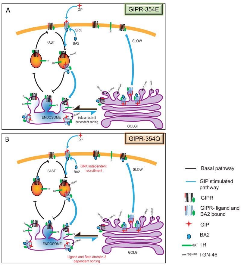
Glucose-dependent insulinotropic polypeptide (GIP) is an incretin hormone involved in nutrient homeostasis. GIP receptor (GIPR) is constitutively internalized and returned to the plasma membrane, atypical behavior for a G-protein-coupled receptor (GPCR). GIP promotes GIPR downregulation from the plasma membrane by inhibiting recycling without affecting internalization. This transient desensitization is achieved by altered intracellular trafficking of activated GIPR. GIP stimulation induces a switch in GIPR recycling from a rapid endosomal to a slow trans-Golgi network (TGN) pathway. GPCR kinases and β-arrestin2 are required for this switch in recycling. A coding sequence variant of GIPR, which has been associated with metabolic alterations, has altered post-activation trafficking characterized by enhanced downregulation and prolonged desensitization. Downregulation of the variant requires β-arrestin2 targeting to the TGN but is independent of GPCR kinases. The single amino acid substitution in the variant biases the receptor to promote GIP-stimulated β-arrestin2 recruitment without receptor phosphorylation, thereby enhancing downregulation.

Keywords: GIP; GIP receptor; GIPR downregulation; GPCR; GPCR trafficking; GRK; beta arrestin; incretin.


Figures

Fig. 1
Fig. 1. βA2 is required for GIP-stimulated GIPR sequestration and slowed recycling
(A) Fixed non-permeabilized adipocytes expressing HA-GIPR-GFP, with or without β-arrestin1 (βA1) or β-arrestin2 (βA2) knockdown (KD). Surface GIPR was stained with Cy3 using anti HA-epitope antibody. Epi fluorescence images. Scale bar: 50μm (B) Quantitation of GIPR plasma membrane to total cell expression (PM-to-Total) distribution in basal and GIP-stimulated (100 nM, 60 min) cells. Adipocytes were electroporated with HA-GIPR-GFP and with no siRNA (WT), βA1, βA2 or βA1+2 siRNA. PM-to-total (Cy3/GFP) were determined as described in materials and methods. Data from individual experiments are normalized to the control cells in basal conditions. Data are averages of 9 independent experiments ± SD., P ≤ 0.05. (C) GIPR internalization in WT adipocytes was measured as described in materials and methods. The slopes, which are the rate constant of internalization, are plotted in panel E. Data are averages ± SD of 9 independent experiments. (D) GIPR internalization experiment in βA2 KD adipocytes. Data are averages ± SD of 9 independent experiments. Each βA2 KD experiment was accompanied by an experiment in WT adipocytes (shown in (C)). (E) Internalization rate constants (Ki) for GIPR internalization in WT or βA2 KD adipocytes. The Ki were calculated as slopes of straight lines from (C) and (D). Data are averages of 9 independent experiments ± SD. (F) GIPR Exocytosis was measured as described in materials and methods. Cell associated Cy3 normalized to GFP was plotted against time. Data are average ± SD of 9 independent experiments. (G) GIPR exocytosis experiment in βA2 KD adipocytes. Data are averages ± SD of 9 independent experiments. Each βA2 KD experiment was accompanied by an experiment in WT adipocytes (shown in (F). (H) Exocytic rate constants (Ke) for GIPR in WT or βA2 KD cells calculated from (F) and (G). The data were fit to a single-phase exponential rise equation. Data are averages of 9 independent experiments ± SD., p<0.05. See also Fig S1.
Fig. 2
Fig. 2. GRKs 2 or 5 are required for GIP-stimulated GIPR sequestration and slowed recycling
(A) Quantitation of GIPR PM-to-Total distribution in basal and GIP-stimulated WT (no siRNA), and GRK KD adipocytes. Data are averages of 5 independent experiments ± SD., p≤0.05. (B) GIPR Exocytosis experiment in WT adipocytes. Data are averages ± SD of 5 independent experiments. (C) GIPR exocytosis in GRK2+5 double KD adipocytes. Data are average ± SD of 5 independent experiments. Each GRK 2+5KD experiment was accompanied by an experiment in WT adipocytes (shown in panel B). (D) Exocytic rate constants (Ke) for GIPR in WT or GRK2+5 KD cells calculated from (B) and (C). The data were fit to a single phase exponential rise equation. Data are averages of 5 independent experiments ± SD., p<0.05. (E) GIPR internalization in WT adipocytes. The internalization rate constants are plotted in panel G. Data are averages ± SD of 7 independent experiments. (F) GIPR internalization in GRK2+5 double KD adipocytes. Data are averages ± SD of 7 independent experiments. Each GRK2+5 double KD was accompanied by an experiment in WT adipocytes (shown in panel E). (G) Internalization rate constants (Ki) for GIPR internalization in WT or βA2 GRK2+5 double KD. The Ki were calculated as slopes of straight lines from (E) and (F). See also Fig S1 and S2.
Fig. 3
Fig. 3. βA2 binding to GIPR is GIP- and GRK2/5-dependent
(A) Immunoblot for co-immunoprecipitated HA-GIPR-GFP blotted for HA-GIPR-GFP and βA2. IP was done with or without GIP stimulation. (Right) Quantification of co immunoprecipitation experiments like that shown in the left panel. βA2 in each lane was normalized to its input and to the GIPR in the IP. Data are averages ± SD of 3 independent experiments., P<0.05. (B) Immunoblot for co-IP of HA-GIPR-GFP in GRK 2+5 KD cells. Blot was done for HA-GIPR-GFP and βA2. (Right) Quantification of co immunoprecipitation experiments like that shown in the left panel. βA2 in each lane was normalized to its input and to the GIPR in the IP.
Fig. 4
Fig. 4. GIPR is localized to TR-containing endosomes in basal conditions and to TGN46-containing membranes upon GIP stimulation
(A) Immunoblot of TGN-46 immunoadsorption from basal and GIP stimulated adipocytes. TGN-46 membranes were immunoisolated using anti-TGN-46 antibody in the absence of detergent. The Input (In), unbound flow-through (FL) and elution (Bd) were run on SDS-PAGE and blotted for TGN markers Syntaxin 6 and mannose-6-phosphate receptor (M-6-P cation independent) and cis-golgi marker GM-130 and for GIPR. (B) Quantification of (A). Relative enrichment of proteins in elution were calculated by normalizing to the flowthrough. (C) Immunoblot of GIPR immunoadsorption from basal and GIP stimulated WT adipocytes. GIPR-GFP membranes were pulled down in absence of detergent. The elution (bound) was run on SDS-PAGE together with input and unbound (FL) and blotted for GIPR, TR and TGN-46. (D) Quantification of (C). Relative amounts of TR or TGN in the elution were calculated by normalizing to input and GIPR in the IP. (E) Quantification of GIP induced GIPR downregulation in syntaxin-6 (Sx-6) knockdown adipocytes. Two different siRNAs were used. Both knockdowns show a decrease in GIPR downregulation showing that it depends on Sx-6. Data are averages ± SD of 3 independent experiments., P<0.05. (F) Exocytosis rate constants for TR and TGN vesicles compared with GIPR in basal and GIP stimulated condition. GIPR ke values are plotted from data in Fig. 1. (G) Schematic representation of GIPR recycling from the ERC (TR-containing endosomes) or TGN compartments with associated rate constants. ki, internalization rate constant, ‘kERC’, rate from the ERC; ‘kTGN’, rate from TGN; and ‘ksort’, GIPR sorting rate from ERC to the TGN. Additional connectivities between these compartments did not significantly improve the performance of this model. (H) Calculation of GIPR sorting rate constant (Ksort) by using equation 1 (supplementary methods) and the model in Fig. 4G. Data from Fig. 1F was used. Inset shows the value of ‘r’ calculated under basal or upon GIP stimulation. (I) GIPR as a fraction of total in different compartments under basal or GIP stimulated conditions. The values were calculated from (F). (J) And (K) Immunoblot of GIPR immunoadsorption from basal and GIP stimulated βA2 KD adipocytes.
Fig. 5
Fig. 5. GIPR intracellular domain is required for GIP induced GIPR sequestration
(A) Amino acid sequence of GIPR predicted cytoplasmic domain (residues 400 to 466). The C-terminal deletion is marked by the red bar. Serines mutated to alanines are noted by arrows. Serines conserved in human, mouse, rat and bovine GIPR are shown in green. The sequence also contains two threonines as potential phosphorylation sites. However, these threonines are not conserved in mammals. (B) Images of GIPRΔ444-466-GFP construct in basal adipocytes. Plasma membrane GIPR was labeled by indirect immunofluorescence with anti-HA/Cy3 secondary antibody of fixed cells. Scale bar: 50μm. (C) Quantification of downregulation of GIPRΔ444-466-GFP constructs upon GIP stimulation. The data are the PM-to-Total of GIP-stimulated normalized to the basal PM-to-Total., P<0.05. (D) GIP-stimulated cAMP production in adipocytes expressing GIPR or GIPRΔ444-466. The endogenous GIPR was knocked down by siRNA such that cAMP production was downstream of the ectopically expressed GIPRs. (E) GIP induced downregulation of the GIPR serine mutants. For each construct, the downregulation has been plotted as PM-to-Total normalized to their basal. The unstimulated level (basal) is shown with a dashed line.
Fig. 6
Fig. 6. Redistribution of the GIPR-Gln354 variant to the TGN is dependent on GIP and βA2 and independent of GRKs
(A) HA-GIPR-Gln354-GFP Immunoadsorption from WT adipocytes and blotted for GIPR, TR and TGN46. (B) Quantification of data from experiments like that shown in panel A. TR and TGN46 in elution were calculated as normalized in total (input) and GIPR in the elution. Data are average ± SD from 3 independent experiments. (C) Effect βA1 or βA2 KD on PM-to-Total distribution of GIPR-Gln354 variant. Data are normalized to their own basal values. Data are averages ± SD of 3 independent experiments., P<0.05. (D) Effect of GRK2+5+6 triple KD on PM-to-Total distribution of GIPR-Gln354 variant. Data are averages ± SD of 3 independent experiments., P<0.05. (E) PM-to-Total distribution of GIPR-Gln354 variant and GIPR-354Gln-Δ444-466 deletion mutant. Data are averages ± SD of 3 independent experiments., P<0.05. (F) Co-immunoprecipitation of βA2 with GIPR-Gln354. (G) Quantification of data from experiments like that shown in panel F. Data are average ± SD from 3 independent experiments. (H) Relative βA2 binding to GIPR and GIPR-354Gln. GIPR and GIPR-354Gln Co-IP were blotted for GIPR and βA2 on the same blot. (I) Quantification of data from experiments like that shown in panel H. Data are average ± SD from 3 independent experiments. (J) GIPR-354Gln-GFP:βA2 binding in GRK2+5 double KD cells. (K) Quantification of data from experiments like that shown in panel H. Data are average ± SD from 3 independent experiments.
Fig. 7
Fig. 7. Schematic of the GIPR trafficking pathway
(A) GIPR is recycled constitutively by the fast TR recycling pathway (black arrows). Upon GIP stimulation, the GIPR is sorted to the slower TGN pathway (blue arrows). This redistribution of recycling pathways results in a dynamic reduction of GIPR from the plasma membrane. The redistribution of GIPR from TR to TGN pathway is regulated by β-arrestin2 (βA2) binding, which in turn is recruited in response to ligand binding and GRK mediated phosphorylation. (B) Downregulation of the GIPR-354Gln variant is by a similar dynamic sequestration mechanism, requiring GIP stimulation and βA2 targeting of the variant from the TR to the TGN recycling pathway. However, βA2 binding to the variant is independent of GRK phosphorylation.

References

    1. Banting G, Ponnambalam S. TGN38 and its orthologues: roles in post-TGN vesicle formation and maintenance of TGN morphology. Biochimica et biophysica acta. 1997;1355:209–217. - PubMed
    1. Bard F, Malhotra V. The formation of TGN-to-plasma-membrane transport carriers. Annu Rev Cell Dev Biol. 2006;22:439–455. - PubMed
    1. Blot V, McGraw TE. GLUT4 is internalized by a cholesterol-dependent nystatin-sensitive mechanism inhibited by insulin. The EMBO journal. 2006;25:5648–5658. - PMC - PubMed
    1. Blot V, McGraw TE. Molecular mechanisms controlling GLUT4 intracellular retention. Mol Biol Cell. 2008;19:3477–3487. - PMC - PubMed
    1. Bruno J, Brumfield A, Chaudhary N, Iaea D, McGraw TE. SEC16A is a RAB10 effector required for insulin-stimulated GLUT4 trafficking in adipocytes. J Cell Biol. 2016;214:61–76. - PMC - PubMed

MeSH terms

Substances

LinkOut - more resources