. 2017 Feb 1;312(2):R172-R188.
doi: 10.1152/ajpregu.00238.2016. Epub 2016 Dec 14.

Kölliker-Fuse nuclei regulate respiratory rhythm variability via a gain-control mechanism

Affiliations

Kölliker-Fuse nuclei regulate respiratory rhythm variability via a gain-control mechanism

Rishi R Dhingra et al. Am J Physiol Regul Integr Comp Physiol. .

Abstract

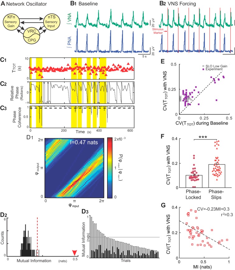
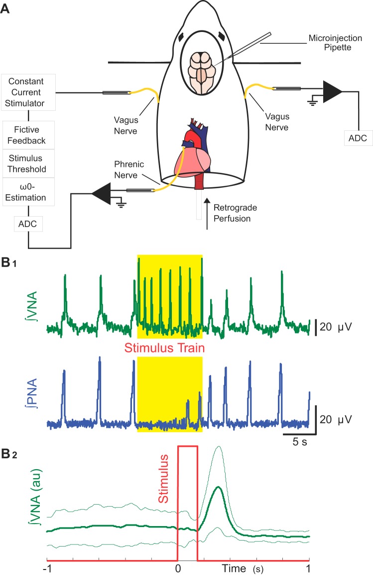
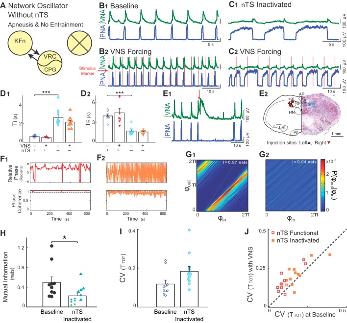
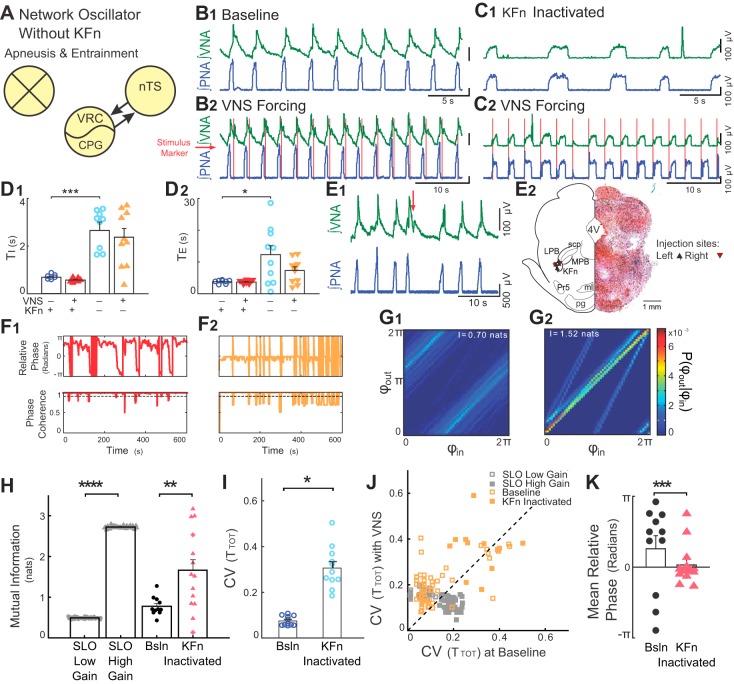
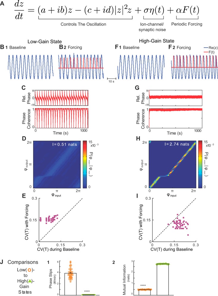
Respiration varies from breath to breath. On the millisecond timescale of spiking, neuronal circuits exhibit variability due to the stochastic properties of ion channels and synapses. Does this fast, microscopic source of variability contribute to the slower, macroscopic variability of the respiratory period? To address this question, we modeled a stochastic oscillator with forcing; then, we tested its predictions experimentally for the respiratory rhythm generated by the in situ perfused preparation during vagal nerve stimulation (VNS). Our simulations identified a relationship among the gain of the input, entrainment strength, and rhythm variability. Specifically, at high gain, the periodic input entrained the oscillator and reduced variability, whereas at low gain, the noise interacted with the input, causing events known as "phase slips", which increased variability on a slow timescale. Experimentally, the in situ preparation behaved like the low-gain model: VNS entrained respiration but exhibited phase slips that increased rhythm variability. Next, we used bilateral muscimol microinjections in discrete respiratory compartments to identify areas involved in VNS gain control. Suppression of activity in the nucleus tractus solitarii occluded both entrainment and amplification of rhythm variability by VNS, confirming that these effects were due to the activation of the Hering-Breuer reflex. Suppressing activity of the Kölliker-Fuse nuclei (KFn) enhanced entrainment and reduced rhythm variability during VNS, consistent with the predictions of the high-gain model. Together, the model and experiments suggest that the KFn regulates respiratory rhythm variability via a gain control mechanism.

Keywords: Hering-Breuer reflex; in situ preparation; respiratory rhythmogenesis; stochastic nonlinear oscillator; vagal nerve stimulation.


Figures

Fig. 3.
Fig. 3.
Rhythmic VNS evokes stable input-output entrainment and increases respiratory rhythm variability in situ. A: schematic model of the Hering-Breuer reflex circuit is shown. The nucleus tractus solitarii (nTS) relays vagal sensory input to the lateral respiratory column, specifically, the ventral respiratory column (VRC) including the central pattern generator (CPG) and Kölliker-Fuse nuclei (KFn). These three regions are connected reciprocally and can be thought of collectively as a single oscillator when analyzing phrenic motor output. B: representative traces of PNA and VNA are shown during baseline (B1) and VNS (B2). B1: at baseline, the in situ arterially perfused preparation generated an in vivo-like pattern of activities, wherein VNA peaked after PNA. B2: during VNS (red lines), each train of VNS evoked bursts of efferent VNA. C: representative plots of the respiratory period (C1, TTOT), instantaneous relative phase (C2, ϕoutputinput) and phase coherence during VNS (C3) are shown. Flat regions in the instantaneous relative phase (C2) indicate periods of perfect phase locking. The phase coherence (C3) measures the mean direction of the relative phase time series in a 20-s sliding window. A phase slip event was defined as a window with a phase coherence less than 0.9 (dashed line in C3) and are highlighted across all three plots. Importantly, the variability in the respiratory period increased selectively during phase slip events. D: joint probability histogram (D1) of the instantaneous phase of the output (PNA; ϕoutput) given the instantaneous phase of the input (VNS stimulus; ϕinput) had strong banding, indicative of stable input-output entrainment. The mutual information for this epoch was 0.47 nats (arrowhead in D2). To determine the significance of the entrainment interaction, the data were bootstrapped by shuffling the interbreath intervals (n = 100) and recomputing the mutual information of the instantaneous phases (D2). The observed value (arrowhead) was greater than the 99% confidence interval of the bootstrapped distribution (dashed line), indicating that the observed input-output entrainment was significant. The mutual information of the instantaneous phases (open bars) and their corresponding 99% confidence intervals (solid bars) was significant for 44 of 45 trials from eight experiments (D3). The distribution of the mutual information of the instantaneous phases derived from simulations of the low-gain SLO model (gray lines) fell slightly above the median of the experimental data set. E: VNS increased the coefficient of variation (CV) of the instantaneous period (purple squares) (Wilcoxon signed-rank test; P = 2.14 × 10⁻⁵). For qualitative comparison, we have also plotted the distribution of CVs from the simulations of the low-gain SLO model (gray squares), which overlap with the experimental distribution. F: for the group of experiential preparations, the CV during phase-locked windows was less than the CV during phase-slip windows, suggesting that the source of the increase in variability during VNS was the phase slip events (***P < 0.001). G: accordingly, the strength of entrainment during rhythmic VNS (as quantified by mutual information) was inversely correlated with the magnitude of respiratory rhythm variability. Together, these results are consistent with the predictions of the low-gain, forced, stochastic SLO model, suggesting that the intact respiratory rhythm-generating network similarly maintains a low-input gain for vagal afferent input.
Fig. 1.
Fig. 1.
Methods to assess whether vagal nerve stimulation (VNS) is sufficient for respiratory rhythm variability and Hering-Breuer input-output entrainment in situ. A: we measured left phrenic (PNA) and right vagal nerve (VNA) activities, while rhythmically forcing the network at its intrinsic oscillation frequency (ω0) via left VNS. B1: threshold amplitude for the electrical stimulus to evoke the Hering-Breuer reflex (HBR) was defined not only by the suppression of inspiratory activity on PNA (bottom), but also by the presence of evoked bursts of efferent VNA from the contralateral vagus nerve (top). B2: stimulus-triggered average of integrated efferent right VNA during a rhythmic VNS trial is shown. The rhythmic VNS (red trace) consistently evoked bursts of efferent VNA (green traces: thick line is the averaged activity; and thin lines, plus and minus standard deviation) at a delay of ~250 ms.
Fig. 4.
Fig. 4.
Suppressing nTS neuronal activity occludes VNS-evoked entrainment and variability amplification. A: we suppressed neuronal activity in discrete components of the Hering-Breuer reflex circuitry via local microinjections of the GABAA receptor agonist, muscimol. Subsequent panels demonstrate that suppression of nTS neuronal activity (marked with an X) occludes both entrainment and variability amplification evoked by rhythmic VNS in situ. B and C: representative traces of PNA and VNA during baseline (B1 and C1) and VNS (B2 and C2) before (B) and after (C) microinjection of muscimol in the nTS. Silencing nTS activity induced apneusis, a prolongation of inspiration. However, a postinspiratory peak in VNA remained, suggesting the presence of an intact three-phase rhythm (Fig. 4C). Furthermore, during VNS, PNA had no consistent phase relation between vagal stimuli and respiration. D: for this experimental group, microinjections of muscimol in the nTS increased inspiratory time (TI) (D1, ***P < 0.001) and decreased expiratory time (TE) (D2, ***P < 0.001). E: respiratory related nTS subnuclei were identified during the experiment by the respiratory responses to local glutamate microinjection (E1) and after the experiment by histologic identification of the injection site (E2). Injections of glutamate (20–50 nl, 10 mM, at red arrow) at the target site evoked VNA and prolonged TE. E2, right: a hemisection counterstained with neutral red is shown indicating the location of a representative muscimol microinjection site marked by pontamine sky blue. E2, left: the locations of the recovered microinjection sites for experiments included in the subsequent analyses are shown. Left and right injection sites are indicated by upward- and downward-pointing triangles, respectively. AP, area postrema; DMNV, dorsal motor nucleus of the vagus; HN, hypoglossal nucleus; LRt, lateral reticular formation; py, pyramidal tract; sol, solitary tract. F: instantaneous relative phase (top) and phase coherence (bottom) are shown before (F1) and after (F2) microinjection of muscimol in the nTS. After suppression of nTS, the respiration did not entrain to VNS. Accordingly, phase coherence was below 0.9, indicating that the underlying instantaneous relative phase window had little directionality. G: joint probability histograms of the instantaneous phase before (G1) and after (G2) microinjection of muscimol in the nTS are shown. Suppression of nTS activity abolished the banding associated with entrainment. H: for the group, bilateral microinjections of muscimol in the nTS decreased the strength of entrainment as measured by the mutual information of the instantaneous phases (*P < 0.05). Note that starred data points indicate VNS stimulation trials that failed the bootstrap test. I: suppression of nTS activity with muscimol tended to increase the variability in TTOT. J: VNS stimulation evoked a significant amplification of CV before (P = 0.004), but not after (P = 0.15) nTS muscimol microinjection. Together, these results confirm that entrainment between VNS and respiration depends on the ability of nTS neurons to relay VNS input to the network. In the context of the model, activity in the nTS is related to the parameter α.
Fig. 5.
Fig. 5.
Suppression of KFn activity evokes high-gain responses to rhythmic VNS. A: suppression of KFn activity spared nTS-CPG connectivity selectively and was sufficient to switch the network state from a low- to high-gain state. B and C: representative traces of PNA and VNA are shown during baseline (B1 and C1) and VNS (B2 and C2) before (B) and after (C) microinjection of muscimol in the KFn. C: suppression of KFn activity increased TI and TE and transformed the respiratory rhythm to a two-phase rhythm without postinspiratory output on vagal efferent fibers. During rhythmic VNS, entrainment strength increased, and the phase angle between VNS and respiration cycle shifted from postinspiratory to preinspiratory coupling. D: for the group, silencing the KFn increased TI (D1, ***P < 0.001) and TE (D2, *P < 0.05). E: respiratory related dorsolateral pontine subnuclei were identified during the experiment by the respiratory responses to local glutamate microinjection (E1) and after the experiment by histological identification of the injection site (E2). E1: injections of glutamate (20–50 nl, 10 mM, at red arrow) at the target site evoked VNA and prolonged TE. E2, right: a representative hemisection counterstained with neutral red shows a KFn-targeted muscimol microinjection site marked by pontamine sky blue. E2, left: locations of the recovered microinjection sites for experiments included in the subsequent analyses are shown. Left and right injection sites are indicated by upward- and downward-pointing triangles, respectively. 4V, fourth ventricle; KFn, Kölliker-Fuse nucleus; LPB, lateral parabrachial nucleus; MPB, medial parabrachial nucleus; ml, medial lemniscus; Pr5, principal sensory of CN V; pg, pontine gray; scp, superior cerebellar peduncle. F: instantaneous relative phase (top) and phase coherence (bottom) are shown before (F1) and after (F2) microinjection of muscimol in the KFn. After suppression of KFn, respiration entrained to VNS with a relative phase near 0 (i.e., the start of inspiration). The reduction in respiratory drive evoked by suppressing KFn activity caused a qualitative change in the desynchronization events, such that they occurred abruptly when respiratory drive was not great enough to evoke an inspiratory burst. G: nonetheless, joint probability histograms of the instantaneous phases before (G1) and after (G2) microinjection of muscimol in the KFn suggest the presence of stronger entrainment after muscimol microinjection. H: for the group, KFn interventions increased the strength entrainment as measured by the mutual information of the instantaneous phases (**P < 0.01), which is consistent with a transition of the state of the network oscillator from low to high gain. For qualitative comparison, we have replotted the effect of increasing gain from the SLO model (****P < 0.001; see Fig. 2). Note that starred data points indicate VNS stimulation trials that failed the bootstrap test. I: at baseline, in the absence of VNS, suppression of KFn activity with muscimol significantly increased the variability in TTOT (*P < 0.05). J: VNS stimulation amplified respiratory rhythm variability before (open orange squares; P < 10–3), but not after (solid orange squares; P = 0.15) KFn muscimol microinjections. For qualitative comparison, we have also plotted the distribution of CVTOT) from simulations of the low- (open gray squares) and high- (solid gray squares) gain SLO models (see Fig. 2). In both model and experimental data, the perturbation (change of gain, or muscimol microinjection) shifted the distribution to lie along the line of identity, suggesting that the amplification of rhythm variability no longer occurred. However, the model failed to capture the increase in rhythm variability evoked by KFn muscimol microinjection. K: suppression of KFn activity shifted the preferred phase during entrainment from a postinspiratory to a preinspiratory relative phase angle (***P = ≪ 0.0001). Together, these observations suggest that activity in the KFn regulates the gain of vagal inputs and is responsible for the amplification of respiratory rhythm variability during VNS forcing.
Fig. 2.
Fig. 2.
A stochastic, forced Stuart-Landau Oscillator (SLO) model predicts that variability during forcing depends on input gain. A: The Stuart-Landau oscillator was modified to include a Gaussian noise term that represents the influence of ion channel gating- and/or synaptic noise on the respiratory oscillation mechanism and a periodic forcing term that represents the influence of periodic vagal input that occurs during mechanical ventilation. B and F: representative outputs of the SLO model (blue traces) in the low- (B) and high- (F) gain states at baseline (B1 and F1) and during rhythmic forcing input (red traces, B2 and F2). C and G: instantaneous relative phase (top) and phase coherence (bottom) of the low- (B) and high- (F) gain states. In the low-gain state, forcing resulted in a coupling pattern that was characterized by epochs of stable coupling interrupted by phase slips qualitatively similar to the experimentally observed coupling pattern (see Fig. 3C). In contrast, in the high-gain state, forcing evoked stronger input-output entrainment without phase slips. D and H: in the low-gain state during forcing (D), the mutual information of the instantaneous phases revealed a significant VNS-evoked entrainment interaction despite the presence of phase slips; whereas, in the high-gain state (H), the mutual information of the instantaneous phases identified a stronger VNS-evoked entrainment interaction. E and I: effect of forcing on rhythm variability also depended on the gain of the model. In the low-gain state (E), forcing increased the variability of the oscillation (P = 9.1 × 10⁻¹⁰). In contrast, in the high-gain state (I), forcing decreased the variability of the oscillation (P = 9.6 × 10⁻¹). J: for the forced, stochastic SLO model, increasing gain reduced the rate of phase slips (****P < 0.0001, J1), and increased the strength of input-output entrainment (****P < < 0.0001, J2). Thus the model predicts that the presence of ion channel- and synaptic-noise sources in the biologic VNS-forced respiratory rhythm generator should generate either stable entrainment characterized by the presence of phase slips and an increase in respiratory rhythm variability, or perfectly phase-locked entrainment without phase slips and a decrease in respiratory rhythm variability depending on the input gain of vagal nerve impulses.

References

    1. Abdala APL, Dutschmann M, Bissonnette JM, Paton JFR. Correction of respiratory disorders in a mouse model of Rett syndrome. Proc Natl Acad Sci USA 107: 18,208–18,213, 2010. doi:10.1073/pnas.1012104107. - DOI - PMC - PubMed
    1. Abdala APP, Bissonnette JM, Newman-Tancredi A. Pinpointing brainstem mechanisms responsible for autonomic dysfunction in Rett syndrome: therapeutic perspectives for 5-HT1A agonists. Front Physiol 5: 205, 2014. doi:10.3389/fphys.2014.00205. - DOI - PMC - PubMed
    1. Abdala AP, Toward MA, Dutschmann M, Bissonnette JM, Paton JFR. Deficiency of GABAergic synaptic inhibition in the Kölliker-Fuse area underlies respiratory dysrhythmia in a mouse model of Rett syndrome. J Physiol 594: 223–237, 2016. doi:10.1113/JP270966. - DOI - PMC - PubMed
    1. Achour L, Letellier C, Cuvelier A, Vérin E, Muir J-F. Asynchrony and cyclic variability in pressure support noninvasive ventilation. Comput Biol Med 37: 1308–1320, 2007. doi:10.1016/j.compbiomed.2006.12.005. - DOI - PubMed
    1. Akoumianaki E, Lyazidi A, Rey N, Matamis D, Perez-Martinez N, Giraud R, Mancebo J, Brochard L, Marie Richard JC. Mechanical ventilation-induced reverse-triggered breaths: a frequently unrecognized form of neuromechanical coupling. Chest 143: 927–938, 2013. doi:10.1378/chest.12-1817. - DOI - PubMed

Publication types

LinkOut - more resources