. 2017 Jan 27;292(4):1374-1384.
doi: 10.1074/jbc.M116.753640. Epub 2016 Dec 14.

Cyclic AMP Inhibits the Activity and Promotes the Acetylation of Acetyl-CoA Synthetase through Competitive Binding to the ATP/AMP Pocket

Affiliations

Cyclic AMP Inhibits the Activity and Promotes the Acetylation of Acetyl-CoA Synthetase through Competitive Binding to the ATP/AMP Pocket

Xiaobiao Han et al. J Biol Chem. .

Abstract

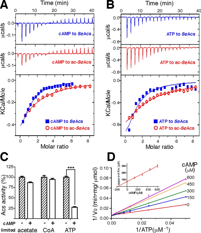
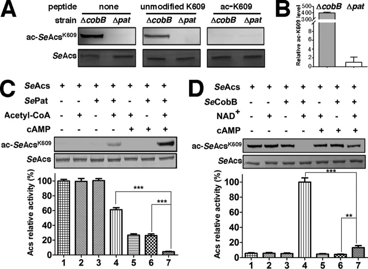
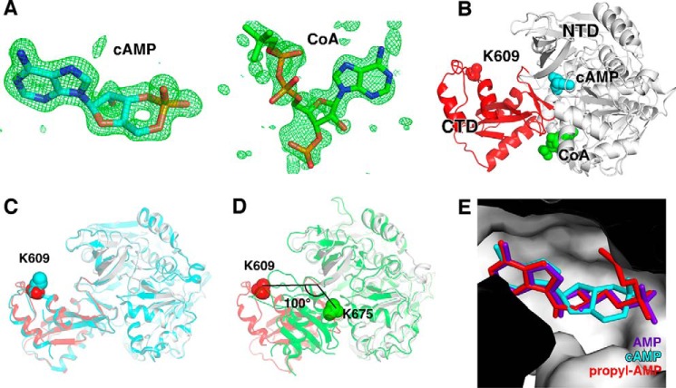
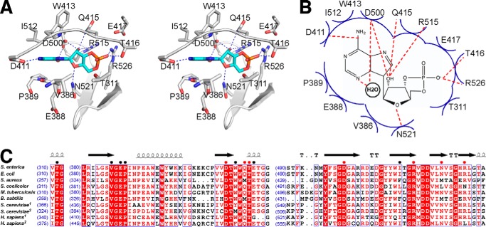
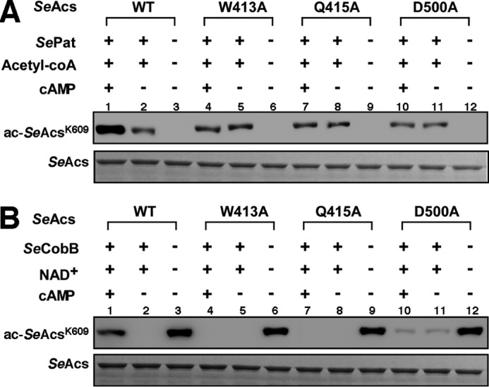
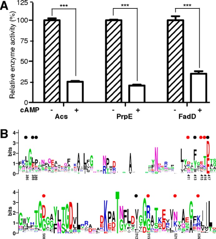
The high-affinity biosynthetic pathway for converting acetate to acetyl-coenzyme A (acetyl-CoA) is catalyzed by the central metabolic enzyme acetyl-coenzyme A synthetase (Acs), which is finely regulated both at the transcriptional level via cyclic AMP (cAMP)-driven trans-activation and at the post-translational level via acetylation inhibition. In this study, we discovered that cAMP directly binds to Salmonella enterica Acs (SeAcs) and inhibits its activity in a substrate-competitive manner. In addition, cAMP binding increases SeAcs acetylation by simultaneously promoting Pat-dependent acetylation and inhibiting CobB-dependent deacetylation, resulting in enhanced SeAcs inhibition. A crystal structure study and site-directed mutagenesis analyses confirmed that cAMP binds to the ATP/AMP pocket of SeAcs, and restrains SeAcs in an open conformation. The cAMP contact residues are well conserved from prokaryotes to eukaryotes, suggesting a general regulatory mechanism of cAMP on Acs.

Keywords: acetyl-CoA synthetase; acetylation; crystal structure; cyclic AMP (cAMP); post-translational modification (PTM).


Figures

FIGURE 1.
FIGURE 1.
cAMP directly binds to SeAcs and inhibits its activity by competing with substrate ATP. A, representative real-time ITC heat change (upper and middle panels) and the integration (lower panel) of each titration of cAMP to SeAcs or acetylated SeAcs (ac-SeAcs). B, representative real-time ITC heat change (upper and middle panels) and the integration (lower panel) of each titration of ATP to SeAcs or ac-SeAcs. C, cAMP inhibits SeAcs at a low concentration of ATP (1 mm CoA, 20 μm ATP, and 20 mm acetate), but not at either a low concentration of acetate (1 mm CoA, 1 mm ATP, and 20 μm acetate), or a low concentration of CoA (20 μm CoA, 1 mm ATP, and 20 mm acetate). The SeAcs activity in the absence of cAMP was treated as 100%. D, the representative Lineweaver-Burk plot shows cAMP inhibits SeAcs by competing with the substrate ATP. The inset panel is a Dixon plot of apparent Km versus cAMP concentration. The Ki of cAMP is estimated from the linear regression. The experiments were repeated in triplicate, and the data are presented as mean ± S.D., ***, p < 0.001.
FIGURE 2.
FIGURE 2.
cAMP promotes acetylation of SeAcs Lys609in vitro. A, the antibody used is specific to acetylated SeAcs Lys609. The same amounts of SeAcs isolated from cobB-null or pat-null S. enterica strains were separated by SDS-PAGE, transferred to the nitrocellulose membrane, then blotted with the antibody specific to the acetylated SeAcs Lys609 in the presence or absence of unmodified peptide (KTRSGKIMRRI), or acetylated peptide (KTRSG(Kac)IMRRI), and detected using an ImageQuant LAS 4000 (GE Healthcare). Coomassie Brilliant Blue staining was used for loading controls. B, semiquantification of the acetylation level of SeAcs Lys609 purified from CobB-null or pat-null S. enterica cells. C, cAMP activates SePat-dependent acetylation and enhances the activity inhibition upon SeAcs. Recombinant SeAcs purified from the pat-null S. enterica strain is treated by acetylation in the presence or absence of cAMP followed by activity measurement and Western blotting detection. The SeAcs activity in the absence of cAMP, acetyl-CoA, and SePat (Lane 1) was treated as 100%. Coomassie Brilliant Blue-stained proteins on SDS-PAGE gels are shown below as a loading control. D, cAMP inhibits SeCob-dependent deacetylation and weakens the activity recovery of SeAcs. Recombinant SeAcs purified from a cobB-null S. enterica strain was treated by deacetylation in the presence or absence of cAMP followed by activity measurement and Western blotting detection. The SeAcs activity (lane 4) was treated as 100%. Each experiment was repeated in triplicate, and the data are presented as mean ± S.D., ***, p < 0.001; **, p < 0.01.
FIGURE 3.
FIGURE 3.
Crystal structure of SeAcs in complex with cAMP and CoA. A, simulated-annealing omit FoFc electron density map (green) contoured at 3.0 σ for cAMP (left) and CoA (right) molecules. Blue, orange, and red represent nitrogen, phosphorus, and oxygen atoms, respectively. Cyan and green represent carbon atoms of cAMP and CoA, respectively. B, overall structure of SeAcs in complex with cAMP (cyan) and CoA (green). White and red ribbons represent the N-terminal and C-terminal domains of SeAcs, respectively; red sphere, Lys609; cyan sphere, cAMP; green sphere, CoA. C, superimposition of our SeAcs-cAMP-CoA crystal structure (red and gray) with the crystal structure of SeAcs complexed with AMP, CoA, and acetate (PDB code 2P2F; cyan). D, superimposition of our SeAcs-cAMP-CoA crystal structure (red and gray) with the crystal structure of yeast Acs in complex with AMP (PDB code 1RY2; green). E, close-up view of the cAMP (cyan) binding pocket with superimposed propyl-AMP (red) from the crystal structure of SeAcs complexed with propyl-AMP, CoA, and acetate (PDB code 1PG4), and with superimposed AMP (purple) from the crystal structure of SeAcs in complex with AMP (PDB code 2P2F).
FIGURE 4.
FIGURE 4.
Detail interactions between SeAcs and cAMP. A, stereo-presentation of detail interactions between SeAcs and cAMP. White ribbon, SeAcs backbone; white stick, SeAcs carbon atoms; cAMP is colored as in panel A. B, schematic presentation of the possible interaction between SeAcs and cAMP. Red dashed lines, H-bonds; blue arcs, Van der Waals interactions. C, sequence alignment of the cAMP-binding pocket in Acs enzymes from different species. The number at the beginning of each line indicates the residue position relative to start of each protein sequence. Residues involved in interactions with cAMP are denoted with colored dots (red, H-bond interaction; black, Van der Waals interaction). Residues conserved in different species are shaded red.
FIGURE 5.
FIGURE 5.
cAMP has no effect on acetylation promotion of mutant SeAcs. A, cAMP activates SePat-dependent acetylation of Lys609 on wild-type SeAcs but has no effect on mutant SeAcs in vitro. B, cAMP inhibits SeCobB-dependent deacetylation of Lys609 on wild-type SeAcs but has no effect on mutant SeAcs in vitro. Top band, acetylation detection by Western blotting using anti-SeAcs Lys609ac antibody; bottom band, Coomassie Brilliant Blue-stained proteins on SDS-PAGE gels are shown as a loading control. Each experiment was independently repeated three times.
FIGURE 6.
FIGURE 6.
cAMP inhibits the activity of other AMP-forming enzymes harboring ATP/AMP binding pockets. A, cAMP inhibits enzymatic activity of SePrpE and SeFadD at a low concentration of ATP (1 mm CoA, 20 μm ATP, and 20 mm acetate), the activities of Acs, PrpE, and FadD in the absence of cAMP were treated as 100%, respectively. Each experiment was independently repeated in triplicate, and the data are presented as mean ± S.D., ***, p < 0.001. B, sequence alignment of 1190 acyl- or aryl-CoA synthetases retrieved from the UniPortKB database (search keyword: EC 6.2.1.- CoA) suggests that the cAMP-binding pocket of Acs is generally conserved in other members of the family. Residues involved in interactions with cAMP are denoted by colored dots as described in the legend to Fig. 4. Residues are numbered as in S. enterica.

References

    1. Pietrocola F., Galluzzi L., Bravo-San Pedro J. M., Madeo F., and Kroemer G. (2015) Acetyl coenzyme A: a central metabolite and second messenger. Cell Metab. 21, 805–821 - PubMed
    1. Wang Q., Zhang Y., Yang C., Xiong H., Lin Y., Yao J., Li H., Xie L., Zhao W., Yao Y., Ning Z. B., Zeng R., Xiong Y., Guan K. L., Zhao S., and Zhao G. P. (2010) Acetylation of metabolic enzymes coordinates carbon source utilization and metabolic flux. Science 327, 1004–1007 - PMC - PubMed
    1. Starai V. J., and Escalante-Semerena J. C. (2004) Acetyl-coenzyme A synthetase (AMP forming). Cell Mol. Life Sci. 61, 2020–2030 - PMC - PubMed
    1. Cederbaum A. I. (2012) Alcohol metabolism. Clin. Liver Dis. 16, 667–685 - PMC - PubMed
    1. Comerford S. A., Huang Z., Du X., Wang Y., Cai L., Witkiewicz A. K., Walters H., Tantawy M. N., Fu A., Manning H. C., Horton J. D., Hammer R. E., McKnight S. L., and Tu B. P. (2014) Acetate dependence of tumors. Cell 159, 1591–1602 - PMC - PubMed

Publication types

MeSH terms

LinkOut - more resources