Review
. 2016 Dec 15;219(Pt 24):3844-3856.
doi: 10.1242/jeb.139899.

Can invertebrates see the e-vector of polarization as a separate modality of light?

Affiliations
Review

Can invertebrates see the e-vector of polarization as a separate modality of light?

Thomas Labhart. J Exp Biol. .

Abstract

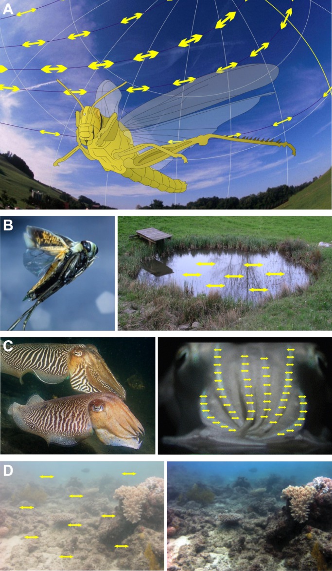
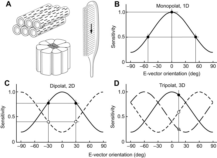
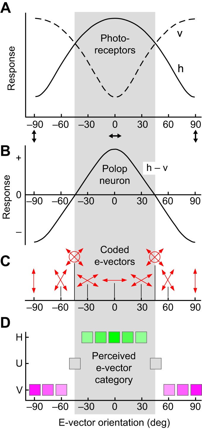
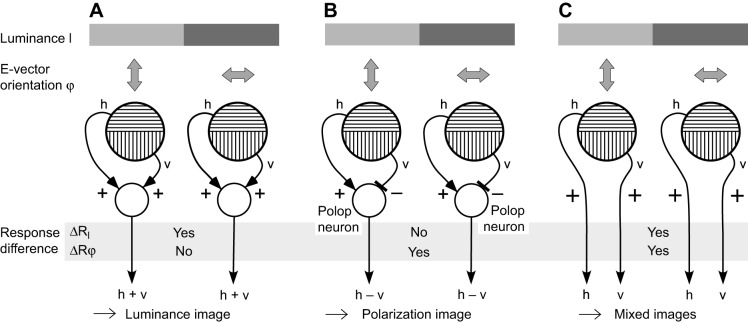
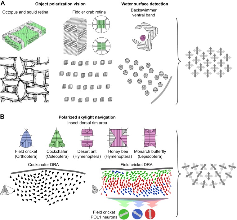
The visual world is rich in linearly polarized light stimuli, which are hidden from the human eye. But many invertebrate species make use of polarized light as a source of valuable visual information. However, exploiting light polarization does not necessarily imply that the electric (e)-vector orientation of polarized light can be perceived as a separate modality of light. In this Review, I address the question of whether invertebrates can detect specific e-vector orientations in a manner similar to that of humans perceiving spectral stimuli as specific hues. To analyze e-vector orientation, the signals of at least three polarization-sensitive sensors (analyzer channels) with different e-vector tuning axes must be compared. The object-based, imaging polarization vision systems of cephalopods and crustaceans, as well as the water-surface detectors of flying backswimmers, use just two analyzer channels. Although this excludes the perception of specific e-vector orientations, a two-channel system does provide a coarse, categoric analysis of polarized light stimuli, comparable to the limited color sense of dichromatic, 'color-blind' humans. The celestial compass of insects employs three or more analyzer channels. However, that compass is multimodal, i.e. e-vector information merges with directional information from other celestial cues, such as the solar azimuth and the spectral gradient in the sky, masking e-vector information. It seems that invertebrate organisms take no interest in the polarization details of visual stimuli, but polarization vision grants more practical benefits, such as improved object detection and visual communication for cephalopods and crustaceans, compass readings to traveling insects, or the alert 'water below!' to water-seeking bugs.

Keywords: Celestial compass; E-vector perception; Invertebrates; Polarization imaging; Polarization vision; Polarization-opponent neurons.


Conflict of interest statement

The author declares no competing or financial interests.

Figures

Fig. 1.
Fig. 1.
Examples of the main functions of polarization vision in invertebrates. (A) Polarization compass: a locust navigating by the polarization pattern of the sky. Image courtesy of Stanley Heinze. (B) Detection of water bodies: a flying backswimmer detects a pond by the horizontal polarization of light reflected from the water surface. Left panel, photograph by Kim Taylor; right panel, photograph by Thomas Labhart. (C,D) Object-based, imaging polarization vision. (C) Cuttlefish recognize the polarized body pattern of conspecifics. Left panel, photograph by Tino Brandt; right panel, photograph modified from Mäthger et al. (2009). (D) Contrast enhancement by reducing the horizontally polarized haze in the water column. The underwater scene (left) was processed using an algorithm that exploits the polarization sensitivity of photoreceptors (Schechner and Karpel, 2004) to produce an enhanced image (right). Note that this is a computer simulation demonstrating the potential gain of visibility afforded by a polarization-sensitive retina; modified from Cronin and Marshall (2011). Yellow double-headed arrows in A–D indicate the dominant e-vector orientation of partial linear polarization. All images used with permission.
Fig. 2.
Fig. 2.
Structural and physiological properties of polarization-sensitive invertebrate photoreceptors. (A) Structural basis of polarization sensitivity in invertebrate photoreceptors. Right, microvilli form the rhabdomere of the photoreceptor. Light travels orthogonal to the microvilli (see arrow). Upper left, the visual pigment molecules with their elongated chromophores (black bars) reside in the microvillar membrane. Alignment of both chromophores and microvilli produces strong polarization sensitivity in the photoreceptor. Lower left, arthropod photoreceptors are grouped to form retinulae. (B–D) E-vector sensitivity functions. (B) A one-dimensional system (monopolat). (C) A two-dimensional system (dipolat) with 90 deg phase-shifted sensitivity functions. (D) A three-dimensional system (tripolat) with 60 deg phase-shifted sensitivity functions. In B–D, polarization sensitivity (PS)=5.0, degree of linear polarization (d)=1.0. A value of 0 deg indicates horizontal e-vector orientation and 90 deg indicates vertical e-vector orientation. Thin straight lines indicate values of selected data points (circular symbols) on the sensitivity and e-vector axes.
Fig. 3.
Fig. 3.
E-vector coding properties of an orthogonal dipolatic (2D) system. (A) E-vector response functions of photoreceptors with horizontal (h) and vertical (v) e-vector tuning axes. (B) E-vector response function of a polarization-opponent (polop) neuron (h−v). (C) E-vector coding by a polop neuron. For a defined degree of polarization (e.g. d=1.0), coding is ambiguous for all e-vectors (double-headed arrows) except for 0 deg and ±90 deg. E-vectors of ±45 deg are also confused with unpolarized light (indicated by circles). For undefined degrees of polarization, not just two but an infinite number of e-vectors can produce the same response in the polop neuron, all appearing identical to a dipolat. (D) The two perceptual categories ‘horizontal’ (H; green symbols) and ‘vertical’ (V; purple symbols) of e-vector orientation perceived by the dipolat. Excitation or inhibition of the polop neuron elicits perception of either H or of V, respectively. Intensity of color indicates perceived saturation. Stimuli of ±45 deg are perceived as unpolarized light (U; gray symbols).
Fig. 4.
Fig. 4.
Effects of interactions between the signals of polarization-sensitive photoreceptors in an orthogonal dipolatic system. (A–C) Two ommatidia view a visual contrast of both luminance (gray bars) and e-vector orientation (double-headed arrows). h and v are the responses of photoreceptors with horizontal and vertical e-vector tuning axes, respectively. Plus and minus signs mark activating and inhibiting connections, respectively. ΔRI and ΔRφ show the difference between the responses of the two ommatidia for luminance contrast and e-vector contrast, respectively. (A) Pooling h and v provides a largely polarization-insensitive luminance image. For receptors with high PS, the pooled signal retains some e-vector dependence because of the logarithmic relation between photon absorption and photoreceptor response (apparent in the log-cos2-shaped e-vector response functions of the photoreceptors of Fig. 3A). (B) Polarization-opponent interaction between h and v by polop neurons produces a luminance-independent polarization image. (C) Keeping h and v separate provides images that are sensitive to both polarization and luminance.
Fig. 5.
Fig. 5.
Overview of the basic sensory principles in invertebrate polarization vision. (A) Two-dimensional (dipolatic) polarization vision. (B) Multidimensional (polypolatic) polarization vision with dipolatic input stages. Upper rows in A and B: rhabdoms of photoreceptors providing polarization vision and their microvilli orientations. Colors indicate the spectral receptor types of the involved photoreceptors; purple indicates UV sensitivity. Note that all input stages are dipolatic. Lower rows in A and B: retinal arrays of ommatidia and photoreceptors. Up is dorsal. Note the characteristic differences between the arrays in A (aligned) and those in B (fan-like). The cricket DRA in B (lower row) is represented in part only. The three cricket POL1 neurons (colored circles in B, lower row) receive input from DRA ommatidia as indicated by the colors; double-headed arrows denote their e-vector tuning axes. Right in A and B: schematic representations of the two types of retinal array, i.e. aligned versus fan-like arrangement of input stages. Crossed double-headed arrows indicate the e-vector tuning axes of dipolatic input stages. Diagrams are not to scale. Cephalopod schematics are modified from Moody and Parriss (1961) and Saidel et al. (1983), with permission. DRA ommatidia are reproduced from Wehner and Labhart (2006), with permission. Other images are after Alkaladi et al. (2013), Labhart et al. (1992, 2001), Schwind (1983) and Waterman and Horch (1966).

References

    1. Alkaladi A., How M. J. and Zeil J. (2013). Systematic variations in microvilli banding patterns along fiddler crab rhabdoms. J. Comp. Physiol. A 199, 99-113. 10.1007/s00359-012-0771-9 - DOI - PubMed
    1. Bernard G. D. and Wehner R. (1977). Functional similarities between polarization vision and color vision. Vision Res. 17, 1019-1028. 10.1016/0042-6989(77)90005-0 - DOI - PubMed
    1. Blum M. and Labhart T. (2000). Photoreceptor visual fields, ommatidial array, and receptor axon projections in the polarisation-sensitive dorsal rim area of the cricket compound eye. J. Comp. Physiol. A 186, 119-128. 10.1007/s003590050012 - DOI - PubMed
    1. Brunner D. and Labhart T. (1987). Behavioural evidence for polarization vision in crickets. Physiol. Entomol. 12, 1-10. 10.1111/j.1365-3032.1987.tb00718.x - DOI
    1. Cartron L., Dickel L., Shashar N. and Darmaillacq A.-S. (2013). Maturation of polarization and luminance contrast sensitivities in cuttlefish (Sepia officinalis). J. Exp. Biol. 216, 2039-2045. 10.1242/jeb.080390 - DOI - PubMed

LinkOut - more resources