. 2017 Feb 14;91(5):e01606-16.
doi: 10.1128/JVI.01606-16. Print 2017 Mar 1.

IRAV (FLJ11286), an Interferon-Stimulated Gene with Antiviral Activity against Dengue Virus, Interacts with MOV10

Affiliations

IRAV (FLJ11286), an Interferon-Stimulated Gene with Antiviral Activity against Dengue Virus, Interacts with MOV10

Corey A Balinsky et al. J Virol. .

Abstract

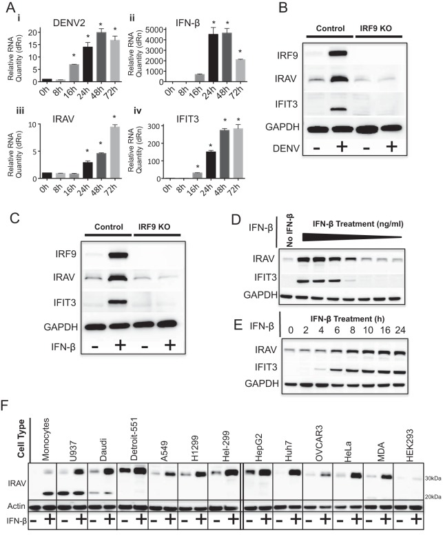
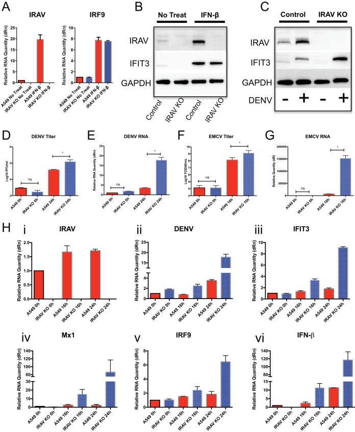
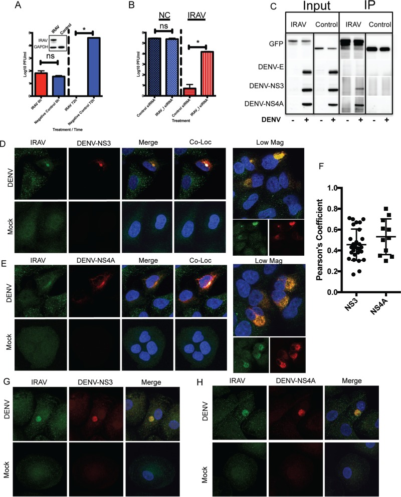
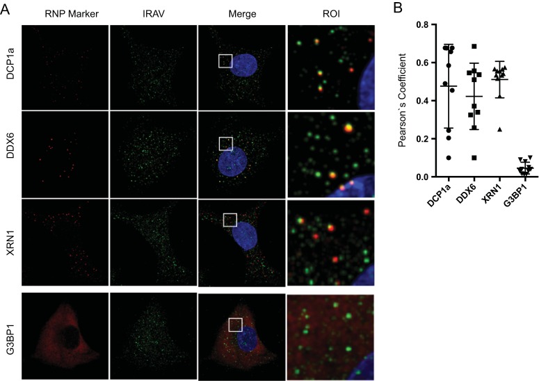
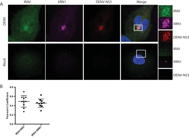
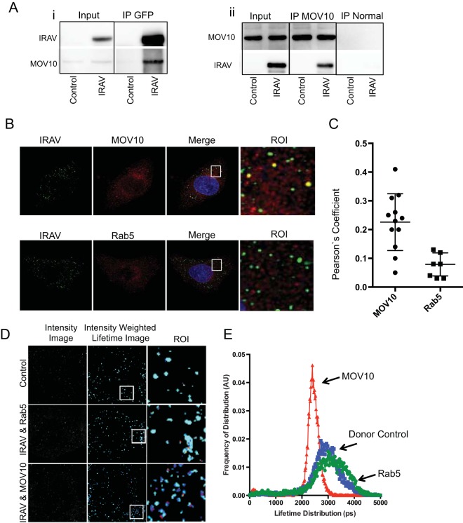
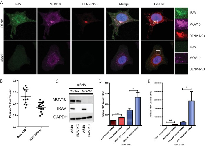
Dengue virus (DENV) is a member of the genus Flavivirus and can cause severe febrile illness. Here, we show that FLJ11286, which we refer to as IRAV, is induced by DENV in an interferon-dependent manner, displays antiviral activity against DENV, and localizes to the DENV replication complex. IRAV is an RNA binding protein and localizes to cytoplasmic processing bodies (P bodies) in uninfected cells, where it interacts with the MOV10 RISC complex RNA helicase, suggesting a role for IRAV in the processing of viral RNA. After DENV infection, IRAV, along with MOV10 and Xrn1, localizes to the DENV replication complex and associates with DENV proteins. Depletion of IRAV or MOV10 results in an increase in viral RNA. These data serve to characterize an interferon-stimulated gene with antiviral activity against DENV, as well as to propose a mechanism of activity involving the processing of viral RNA. IMPORTANCE Dengue virus, a member of the family Flaviviridae, can result in a life-threatening illness and has a significant impact on global health. Dengue virus has been shown to be particularly sensitive to the effects of type I interferon; however, little is known about the mechanisms by which interferon-stimulated genes function to inhibit viral replication. A better understanding of the interferon-mediated antiviral response to dengue virus may aid in the development of novel therapeutics. Here, we examine the influence of the interferon-stimulated gene IRAV (FLJ11286) on dengue virus replication. We show that IRAV associates with P bodies in uninfected cells and with the dengue virus replication complex after infection. IRAV also interacts with MOV10, depletion of which is associated with increased viral replication. Our results provide insight into a newly identified antiviral gene, as well as broadening our understanding of the innate immune response to dengue virus infection.

Keywords: C19orf66; FLJ11286; IRAV; MOV10; P body; UPF0515; dengue; flavivirus; interferon.


Figures

FIG 1
FIG 1
IRAV is an interferon-stimulated gene. (A) qRT-PCR analysis of RNA expression in A549 cells after DENV infection at 0, 8, 16, 24, 48, and 72 h postinfection. Cells were infected with DENV at an MOI of 0.1 for 1 h, and samples were collected at time zero and the indicated time points. The samples were analyzed by qRT-PCR for DENV RNA (i), as well as for expression of IFN-β (ii), IRAV (iii), and the ISG IFIT3 (iv). The samples were normalized to the HPRT housekeeping gene, and the change in expression was calculated relative to time zero. The error bars represent standard deviations. *, P < 0.05. (B) Western blot analysis of IRAV expression in control A549 cells or IRF9 KO cells after DENV infection (72 h). Uninfected (−) and DENV-infected (+) A549 or IRF9 KO cells were analyzed by Western blotting for IRF9, IRAV, or IFIT3. GAPDH was used as a loading control. (C) Western blot analysis of IRAV expression in control A549 or IRF9 KO cells after treatment with IFN-β. The cells were either left untreated (−) or treated with IFN-β (+) for 16 h, followed by Western blot analysis for expression of IRF9, IRAV, or IFIT3. GAPDH was used as a loading control. (D) Western blot analysis of IRAV expression in HeLa cells after treatment with 10-fold dilutions of IFN-β (30 to 0.00003 ng/ml) for 16 h. Samples were examined by Western blotting for expression of IRAV and IFIT3. GAPDH was used as a loading control. (E) Western blot analysis of IRAV expression in HeLa cells after treatment with IFN-β (3 ng/ml) at the indicated time points. Samples were examined by Western blotting for expression of IRAV and IFIT3. GAPDH was used as a loading control. (F) Western blot analysis of IRAV expression in various cell lines. The indicated cell lines were either left untreated (−) or treated with IFN-β (+) (10 ng/ml) for 16 h, followed by Western blot analysis for IRAV. Actin was used as a loading control.
FIG 2
FIG 2
Knockout of IRAV results in enhanced replication of DENV and EMCV. (A) A549 cells or IRAV KO cells were left untreated or treated with IFN-β for 16 h, followed by qRT-PCR analysis of RNA expression of IRAV or IRF9. Samples were normalized to the HPRT housekeeping gene, and the change in expression was calculated relative to time zero. (B) Western blot analysis of A549 control or IRAV KO cells after IFN treatment. The cells were either left untreated or treated with IFN-β for 16 h, followed by Western blot analysis for IRAV or IFIT3. GAPDH was used as a loading control. (C) Western blot analysis of A549 control or IRAV KO cells after DENV infection. The cells were either left untreated (−) or infected with DENV for 72 h (+), followed by Western blotting for IRAV or IFIT3. GAPDH was used as a loading control. (D) A549 or IRAV KO cells were infected with DENV, and samples were collected at time zero and 24 h, followed by titration using the plaque assay method. (E) A549 or IRAV KO cells were infected with DENV, and samples were collected at time zero and 24 h, followed by qRT-PCR analysis of DENV RNA. (F) A549 or IRAV KO cells were infected with EMCV, and samples were collected at time zero and 16 h, followed by titration using the 50% tissue culture infective dose (TCID50) method. (G) A549 or IRAV KO cells were infected with EMCV, and samples were collected at time zero and 16 h, followed by qRT-PCR analysis of EMCV RNA. (H) qRT-PCR analysis of A549 or IRAV KO cells infected with DENV. Samples were collected at time zero and 16 h and 24 h postinfection and analyzed for IRAV (i) and DENV RNA (ii), as well as for expression of IFIT3 (iii), Mx1 (iv), IRF9 (v), and IFN-β (vi). Samples were normalized to the HPRT housekeeping gene, and the change in expression was calculated relative to time zero. The error bars represent standard deviations. *, P < 0.05; ns, not significant.
FIG 3
FIG 3
IRAV associates with the DENV replication complex. (A) HEK293 cells stably expressing IRAV or a negative control were infected with DENV at an MOI of 0.1, and samples were collected at time zero and 72 h postinfection, followed by titration using the plaque assay method. The inset represents expression of IRAV in transfected HEK293 cells compared to the negative control, as determined by Western blot analysis. GAPDH was used as a loading control. (B) HEK293 cells stably expressing IRAV or negative-control (NC) cells were treated with either negative-control siRNA or an siRNA specific to IRAV (IRAV_I). The cells were then infected with DENV for 72 h, followed by titration on Vero cells. (C) IRAV coimmunoprecipitates with DENV proteins. HEK293 cells were either left uninfected (−) or infected with DENV (+) for 48 h, followed by transfection with a plasmid expressing GFP-IRAV (IRAV) or GFP-CAT (Control) for an additional 48 h. The cell lysates were then collected, and co-IP experiments were performed using antibodies to GFP. Input and IP samples were then analyzed by Western blotting for the presence of DENV envelope (DENV-E), DENV NS3, or DENV NS4A protein. (D) Confocal microscopy of IRAV colocalized with replication complexes in DENV-infected or mock-infected A549 cells. Green, IRAV; red, DENV-NS3. The nucleus was stained with DAPI (blue). Colocalization between IRAV and DENV-NS3 is shown in white. Low Mag, a lower-magnification field showing a cluster of infected cells. (E) Confocal microscopy of IRAV colocalized with replication complexes in DENV-infected or mock-infected A549 cells. Green, IRAV; red, DENV NS4A. The nucleus was stained with DAPI (blue). Colocalization between IRAV and DENV NS4A is shown in white. (F) Colocalization coefficients of IRAV and DENV NS3 (NS3) or DENV NS4A (NS4A) in DENV-infected A549 cells as determined by Pearson's linear correlation coefficient. (G) Confocal microscopy of IRAV colocalized with replication complexes in DENV-infected or mock-infected monocyte-derived macrophages. Green, IRAV; red, DENV-NS3. The nucleus was stained with DAPI (blue). (H) Confocal microscopy of IRAV colocalized with replication complexes in DENV-infected or mock-infected monocyte-derived macrophages. Green, IRAV; red, DENV NS4A. The nucleus was stained with DAPI (blue). The error bars represent standard deviations.
FIG 4
FIG 4
IRAV interacts with RNA binding proteins. (A) Gene ontology analysis of IRAV interaction partners as identified by co-IP of GFP-IRAV from DENV-infected HEK293 cells, followed by MS analysis. Gene ontology analysis was performed using the PANTHER classification system (http://www.pantherdb.org), based on the protein class. (B) FP assays were performed using various concentrations of recombinant IRAV and FAM-labeled 20-mer single-stranded or double-stranded DNA or RNA oligomers. Samples were run in triplicate, and FP was measured on a Hidex sense microplate reader (Turku, Finland). (C) Co-IP experiments performed on HEK293 cells left untreated (−) or infected with DENV (+). The cells were transfected with expression vectors for GFP-IRAV (IRAV) or GFP-CAT (Control), followed by IP with an antibody for GFP. Western blotting was performed for IRAV putative interaction partners MOV10, UPF1, and HuR. (D) Co-IP experiments were performed on HEK293 cells after overexpression of GFP-IRAV (IRAV) or GFP-CAT (Control). Cell lysates were either left untreated (−) or treated with RNase A (+), followed by IP with an antibody for GFP. Western blots were then performed for IRAV putative interaction partners MOV10, UPF1, and HuR.
FIG 5
FIG 5
IRAV associates with P bodies in IFN-β-treated cells. (A) Confocal microscopy of IRAV; P body markers DCP1a, DDX6, and XRN1; and the stress granule marker G3BP1a in A549 cells after treatment with IFN-β (10 ng/ml) for 16 h. Green, IRAV; red, RNP markers. The nucleus was stained with DAPI (blue). Regions of interest (ROI) are boxed in white. (B) Colocalization coefficients of IRAV with DCP1a, DDX6, XRN1, and G3BP1 as determined by Pearson's linear correlation coefficient. The error bars represent standard deviations.
FIG 6
FIG 6
IRAV relocalizes to the replication complex after DENV infection. (A) Confocal microscopy of XRN1 colocalized with IRAV and DENV NS3 at the replication complex in DENV-infected or mock-infected A549 cells. Green, IRAV; magenta, XRN1; red, DENV NS3. The nucleus was stained with DAPI (blue). Colocalization between IRAV, XRN1, and DENV NS3 is shown in white. Regions of interest (ROI) are boxed in white. (B) Colocalization coefficients of IRAV and DENV NS3 or IRAV and XRN1 in DENV-infected A549 cells as determined by Pearson's linear correlation coefficient, demonstrating colocalization between IRAV and both XRN1 and DENV NS3 in DENV-infected cells. The error bars represent standard deviations.
FIG 7
FIG 7
IRAV interacts with MOV10. (A) Co-IP experiments performed on HEK293 cells transfected with expression vectors for GFP-IRAV (IRAV) or GFP-CAT (Control), followed by IP with either an antibody for GFP (i) or endogenous MOV10 (ii). Normal IgG was used as a negative control for endogenous MOV10. Western blots were then performed for IRAV or MOV10. (B) Confocal microscopy of IRAV and MOV10 or Rab5 in IFN-β-treated A549 cells. Green, IRAV; red, MOV10 or Rab5. The nucleus was stained with DAPI (blue). ROI are boxed in white. (C) Colocalization coefficients of IRAV and MOV10 or Rab5 in IFN-β-treated A549 cells as determined by Pearson's linear correlation coefficient. (D) Representative image of FRET-by-FLIM analysis of IRAV interactions with MOV10 or Rab5 in IFN-β-treated A549 cells, showing the image intensity, intensity-weighted lifetime, and selected ROI. (E) Lifetime distribution plot versus frequency of distribution, in arbitrary units (AU), of FRET-by-FLIM analysis of IRAV (donor control), IRAV and MOV10, or IRAV and Rab5 in IFN-β-treated A549 cells. The plot was constructed using data from 10 images each, collected from two independent experiments. IRAV interactions with MOV10 are shown in red, Rab5 in green, and the donor control in blue. The error bars represent standard deviations.
FIG 8
FIG 8
MOV10 localizes to the DENV replication complex and restricts viral replication. (A) Confocal microscopy of MOV10 colocalized with IRAV and DENV NS3 at the replication complex in DENV-infected or mock-infected A549 cells. Green, IRAV; magenta, MOV10; red, DENV NS3. The nucleus was stained with DAPI (blue). Colocalization between IRAV, MOV10, and DENV NS3 is shown in white. ROI are boxed in white. (B) Colocalization coefficients of IRAV and DENV NS3 or IRAV and MOV10 in DENV-infected A549 cells as determined by Pearson's linear correlation coefficient, demonstrating colocalization between IRAV and both MOV10 and DENV NS3. (C) Western blot analysis of siRNA-mediated knockdown of MOV10 in A549 or IRAV KO cells. The cells were treated with a gene-specific siRNA for MOV10 (Qiagen Hs_MOV10_5 FlexiTube siRNA) or a negative-control siRNA for 48 h, followed by Western blot analysis for MOV10 or IRAV. GAPDH was used as a loading control. (D) qRT-PCR analysis of A549 or IRAV KO cells treated with either negative-control siRNA or gene-specific MOV10 siRNA for 48 h. The cells were then infected with DENV (MOI, 0.1) for 24 h and analyzed for DENV RNA. Samples were normalized to the HPRT housekeeping gene, and the change in expression was calculated relative to time zero. (E) qRT-PCR analysis of A549 or IRAV KO cells treated with either negative-control siRNA or gene-specific MOV10 siRNA for 48 h. The cells were then infected with EMCV (MOI, 0.001) for 24 h and analyzed for EMCV RNA. Samples were normalized to the HPRT housekeeping gene, and the change in expression was calculated relative to time zero. The error bars represent standard deviations. *, P < 0.05; ns, not significant.

References

    1. Bhatt S, Gething PW, Brady OJ, Messina JP, Farlow AW, Moyes CL, Drake JM, Brownstein JS, Hoen AG, Sankoh O, Myers MF, George DB, Jaenisch T, Wint GRW, Simmons CP, Scott TW, Farrar JJ, Hay SI. 2013. The global distribution and burden of dengue. Nature 496:504–507. doi:10.1038/nature12060. - DOI - PMC - PubMed
    1. Guzman MG, Halstead SB, Artsob H, Buchy P, Farrar J, Gubler DJ, Hunsperger E, Kroeger A, Margolis HS, Martínez E, Nathan MB, Pelegrino JL, Simmons C, Yoksan S, Peeling RW. 2010. Dengue: a continuing global threat. Nat Rev Microbiol 8:S7–S16. doi:10.1038/nrmicro2460. - DOI - PMC - PubMed
    1. Kyle JL, Harris E. 2008. Global spread and persistence of dengue. Annu Rev Microbiol 62:71–92. doi:10.1146/annurev.micro.62.081307.163005. - DOI - PubMed
    1. WHO. 2006. WHO scientific working group report on dengue; reference number TDR/SWG/08. WHO, Geneva, Switzerland.
    1. Schneider WM, Chevillotte MD, Rice CM. 2014. Interferon-stimulated genes: a complex web of host defenses. Annu Rev Immunol 32:513–545. doi:10.1146/annurev-immunol-032713-120231. - DOI - PMC - PubMed