

Correlation of Intraoperative Frozen Section Report and Histopathological Diagnosis of Central Nervous System Tumors - A Six-Year Retrospective Study
- PMID: 27974956
- PMCID: PMC5099397
- DOI: 10.5001/omj.2016.84
Correlation of Intraoperative Frozen Section Report and Histopathological Diagnosis of Central Nervous System Tumors - A Six-Year Retrospective Study
Abstract
Objectives: To evaluate the degree of agreement between the intraoperative frozen section (FS) reporting of central nervous system (CNS) tumors and final histopathological diagnosis based on permanent paraffin section.
Methods: All CNS tumor cases with a diagnosis at FS and subsequent permanent section (n = 261) taken from 2007 to 2012 were retrospectively reviewed. Twenty percent of FS were double-checked by a senior pathologist as part of the study and the intraobserver agreement between the pathologist and the agreement between final report, and initial FS report was estimated by the intraclass correlation coefficient (ICC).
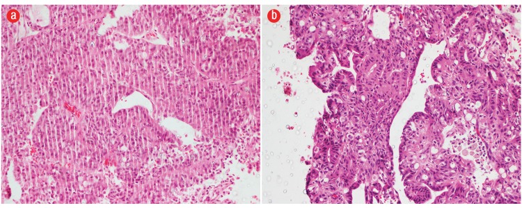
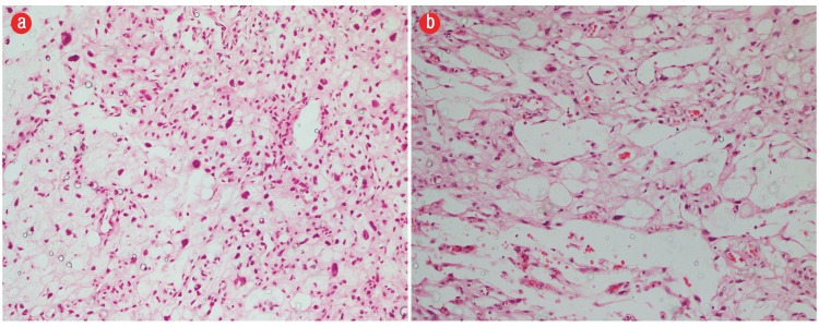
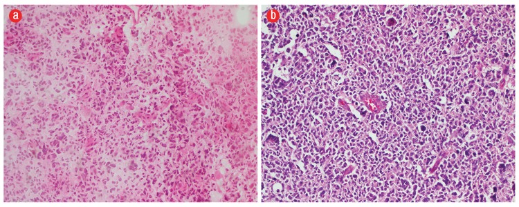
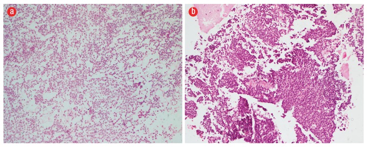
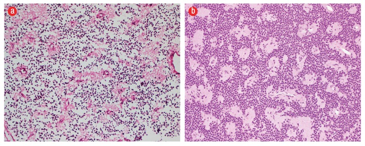
Results: A total of 261 cases were reviewed. The most common diagnosis was glioblastoma (grade IV) and meningioma (grade I-II) forming 45.6% of cases. Fifty-three cases were subjected to intraobserver agreement of histological diagnosis. There was nearly perfect intraobserver agreement on histopathology (ICC = 0.9). Out of 261 cases, 224 cases showed a strong agreement between the FS diagnosis and final histological diagnosis (ICC = 0.747). A discrepancy between the FS and final diagnosis were found in eight cases. The disagreement did not relate to any specific tumor type. However, in three cases, the discrepancy was in the grading of the glioma. In 29 cases, a definite opinion could not be given on FS as the samples examined were nonrepresentative.
Conclusions: Histopathological slides classified by World Health Organization criteria of CNS tumors had excellent intraobserver agreement. Our results show a moderate to high degree of agreement in the intraoperative diagnosis of CNS lesions using FS. However, there are limitations, and some lesions are a diagnostic challenge. There is a need to improve our diagnostic skills and knowledge of possible errors and establish better communication with neurosurgeons.
Keywords: Central Nervous System; Frozen Sections; Intraoperative Procedures.
Figures
References
-
- Plesec TP, Prayson RA. Frozen section discrepancy in the evaluation of central nervous system tumors. Arch Pathol Lab Med 2007. Oct;131(10):1532-1540. - PubMed
-
- Yachnis AT. Intraoperative consultation for nervous system lesions. Semin Diagn Pathol 2002. Nov;19(4):192-206. - PubMed
-
- Ud Din N, Memon A, Idress R, Ahmad Z, Hasan S. Central nervous system lesions: correlation of intraoperative and final diagnoses, six year experience at a referral centre in a developing country, Pakistan. Asian Pac J Cancer Prev 2011;12(6):1435-1437. - PubMed
LinkOut - more resources
Full Text Sources
Other Literature Sources