Review
. 2017 Feb;147(2):149-174.
doi: 10.1007/s00418-016-1520-x. Epub 2016 Dec 14.

Sialylation of N-glycans: mechanism, cellular compartmentalization and function

Affiliations
Review

Sialylation of N-glycans: mechanism, cellular compartmentalization and function

Gaurang P Bhide et al. Histochem Cell Biol. 2017 Feb.

Abstract

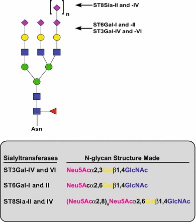
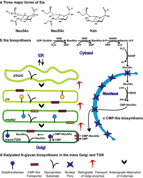
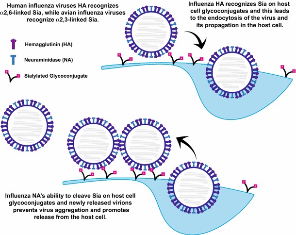
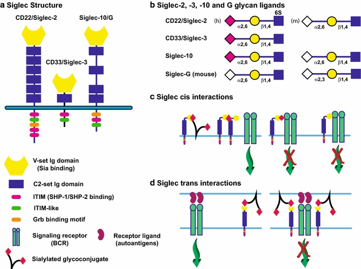
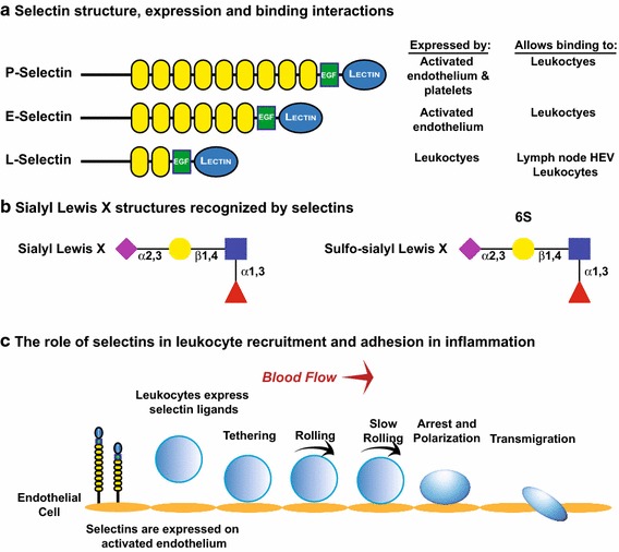
Sialylated N-glycans play essential roles in the immune system, pathogen recognition and cancer. This review approaches the sialylation of N-glycans from three perspectives. The first section focuses on the sialyltransferases that add sialic acid to N-glycans. Included in the discussion is a description of these enzymes' glycan acceptors, conserved domain organization and sequences, molecular structure and catalytic mechanism. In addition, we discuss the protein interactions underlying the polysialylation of a select group of adhesion and signaling molecules. In the second section, the biosynthesis of sialic acid, CMP-sialic acid and sialylated N-glycans is discussed, with a special emphasis on the compartmentalization of these processes in the mammalian cell. The sequences and mechanisms maintaining the sialyltransferases and other glycosylation enzymes in the Golgi are also reviewed. In the final section, we have chosen to discuss processes in which sialylated glycans, both N- and O-linked, play a role. The first part of this section focuses on sialic acid-binding proteins including viral hemagglutinins, Siglecs and selectins. In the second half of this section, we comment on the role of sialylated N-glycans in cancer, including the roles of β1-integrin and Fas receptor N-glycan sialylation in cancer cell survival and drug resistance, and the role of these sialylated proteins and polysialic acid in cancer metastasis.

Keywords: Golgi; Polysialic acid; Selectins; Sialic acid; Sialyltransferase; Siglecs.


Figures

Fig. 1
Fig. 1
N-glycan structures synthesized by α2,3-, α2,6- and α2,8-sialyltransferases. In this review, we have focused on the sialyltransferases that add Sia to N-glycans. The ST3Gal enzymes may also modify O-glycans and glycolipids, while the role of ST6Gal-II in modifying protein-bound N-glycans has not been unequivocally demonstrated. The polysialyltransferases, ST8Sia-II and ST8Sia-IV, synthesize polySia chains of 8 to greater than 400 units long on a preexisting Sia that is many times α2,6-linked. Shown is a triantennary N-glycan as a model and does not imply that the activity of these enzymes in any way is restricted to this type of N-glycan. Pink diamond, Neu5Ac; yellow circle, galactose (Gal); blue square, N-acetylglucosamine (GlcNAc); green circle, mannose (Man); red triangle, fucose (Fuc)
Fig. 2
Fig. 2
Sialyltransferase conserved sequences, domain structure and sequences involved in Golgi localization. a The positions of the conserved sequences found in all STs, the sialylmotifs, are shown using a generic polyST structure. SML, large sialylmotif; SMS, small sialylmotif; 3, motif 3; VS (or SMVS in text), very small sialylmotif. These sequences are involved in glycan substrate and CMP-Sia donor binding as well as catalysis. A key catalytic His residue mentioned in the text is the first residue in the SMVS. Also shown are two sequences conserved in the polySTs, the polybasic region (PBR) and polysialyltransferase domain (PSTD), which are involved in substrate recognition (via protein–protein interactions—PBR) and that are predicted to form a basic surface or groove for the growing polySia chain (PBR and PSTD). The disulfide formed between the SML and the SMS is conserved across all STs, while the disulfide bond linking the C-terminus is essential for the polySTs. b Sialyltransferase domain structure (left) and Golgi localization sequences (right). STs are type II membrane proteins with short, N-terminal cytoplasmic tails followed by relatively short transmembrane (TM) regions, proteolytically sensitive stem regions followed by large catalytic domains that contain the sialylmotifs and face the Golgi lumen. Golgi glycosylation enzymes are localized by multiple mechanisms. Shown is a summary of sequences and mechanisms involved in Golgi localization
Fig. 3
Fig. 3
The protein-specific polysialylation of NCAM and NRP-2. The polysialylation of NCAM and NRP-2 requires that the polySTs recognize and bind an acidic patch or surface on the domain adjacent to the domain or region that carries the glycans to be polysialylated. NCAM consists of five immunoglobulin domains (Ig1–Ig5) and two fibronectin type III repeats (FN1 and 2). NRP-2 consists of two complement homology (CUB) domains, two coagulation factor V/VIII homology (F5/8) domains and one Meprin-A5 protein-μ tyrosine phosphatase (MAM) domain. N-glycans are shown as Vs, O-glycans are shown as horizontal lines, and polySia chains are shown in red. For NCAM polysialylation, either ST8Sia-II or ST8Sia-IV recognize and dock on an acidic patch on the first fibronectin type III repeat (FN1) to allow the polysialylation of two N-glycans in the adjacent Ig5 domain (left). Likewise, for NRP-2, acidic residues of the MAM domain are required for ST8Sia-IV to polysialylate O-glycans in the adjacent linker region between the MAM domain and the second F5/8 domain (F5/8 #2)
Fig. 4
Fig. 4
Sialic acid structure and the compartmentation of Sia, CMP-Sia and sialylated N-glycan biosynthesis. a Three major forms of Sia are shown. b The pathway for Neu5Ac biosynthesis in the cytosol. GNE, UDP-GlcNAc 2-epimerase/ManNAc Kinase; NANS, N-acetylneuraminic acid synthase; NANP, N-acetylneuraminic acid phosphatase. c The pathway for CMP-Sia biosynthesis in the nucleus is shown. Both Neu5Ac and CMP-Neu5Ac are small enough to flow into and out of the nucleus through nuclear pores. CMAS, CMP-sialic acid synthetase. d The pathway for sialylated N-glycan biosynthesis in the trans Golgi and TGN. According to the cisternal maturation model, Golgi enzymes are localized/retained in the Golgi by continuous retrograde transport in COPI-coated vesicles or tubules and cisternae-containing cargo proteins are “matured” by the sequential introduction of glycosylation enzymes. The sialyltransferase reaction in the Golgi trans cisternae and TGN is highlighted. CMP-Sia, like other nucleotide sugar donors, is transported into the Golgi by a specific CMP-Sia transporter (CST). It is then used in the transferase reaction with the release of CMP which is transported to the cytosol by the CST in exchange for CMP-Sia
Fig. 5
Fig. 5
Influenza virus hemagglutinin and neuraminidase recognize Sia residues on host cell glycoconjugates. The hemagglutinin (HA) proteins on the influenza virus membrane recognize Sia residues on host cell surface glycoconjugates. This interaction allows the endocytosis of the virus and its propagation inside the host cell. Release of the newly made virions from the host cell can be hampered by HA–Sia interactions between the host cell surface glycoconjugates and the virus. Sia-mediated interactions between viruses can also cause aggregation at the host cell surface. The viral neuraminidase (NA) cleaves Sia allowing the release of the virus from the host cell and the dispersion of aggregates. Many influenza drugs are designed to block the activity of the NA. Influenza viruses that infect humans recognize α2,6-linked Sia, while influenza viruses that recognize avian species recognize α2,3-linked Sia
Fig. 6
Fig. 6
Siglecs and their role in the regulation of the immune response. a Siglecs share a common structure with an N-terminal V-set Ig domain that recognizes Sia, followed by a variable number of C2-set Ig domains, a transmembrane region and cytoplasmic tail. The cytoplasmic tail of Siglecs contains a number of binding sequences. Inhibitory Siglecs contain immunoreceptor tyrosine-based inhibitory motif (ITIM) sequences that allow SHP-1 and SHP-2 binding and inhibition of signaling, in addition to other signaling molecule-binding sequences. Activating Siglecs (not shown) possess a basic residue in their TM regions that allow the binding of DAP12, an immunoreceptor tyrosine-based activation motif (ITAM) adaptor protein that promotes the activation of signaling. Examples of Siglecs that bind sialylated N-glycan structures are shown. b The sialylated ligands recognized by selected Siglecs are shown. Siglec-10 (human) and Siglec-G (mouse) are considered orthologs; however, the human protein recognizes only α2,6-linked Neu5Ac (pink diamond) or Neu5Gc (white diamond) structures, while the mouse Siglec recognizes both α2,3- and α2,6-linked Sia in the Neu5Gc form. c Siglecs can bind ligands in cis. An inhibitory Siglec can be sequestered away from an activating receptor by binding to other sialylated ligands, including itself (left). Conversely, binding to an activating receptor directly or via Sia residues on its glycans can inhibit its signaling (right). d Siglecs can bind ligands in trans. These interactions can either sequester the Siglec away from an activating receptor (left), or if sialylated ligands are close to the ligands for the activating receptor, this interaction may enhance the ability of the Siglec to inhibit the activating receptor (right). Please see Varki and Crocker (2009), Macauley et al. (2014) for more details on Siglec structure and function
Fig. 7
Fig. 7
Selectins mediate binding to glycans capped with sialyl Lewis X structures to mediate cells interactions in inflammation, lymphocyte homing and metastasis. a Structures of P-, E- and L-selectins, cells on which they are expressed, and the binding they mediate. b Sialyl Lewis X and sulfo-sialyl Lewis X structures. Note that while all selectins recognize sialyl Lewis X, sulfo-sialyl Lewis X is primarily expressed by peripheral lymph node addressins (PNAds) recognized by L-selectin. c Selectin function in leukocyte recruitment and adhesion in inflammation (McEver and Zhu 2010). P- and E-selectins are expressed by activated endothelium and mediate recruitment, tethering and initial stages of leukocyte rolling on the endothelium. Signaling through selectin ligands on the leukocytes stimulates a conformational change in integrins expressed by leukocytes, leading to weak binding of their receptors on the endothelium and slowing their rolling. Release of chemokines from endothelium stimulates signaling from chemokine receptors on the leukocyte membrane, and these signals stimulate the conversion of integrins to forms with high affinity for ligands, and these high-affinity interactions lead to leukocyte arrest. Ultimately, the leukocyte migrates through the endothelium to the site of inflammation

References

    1. Abe M, Noda Y, Adachi H, Yoda K. Localization of GDP-mannose transporter in the Golgi requires retrieval to the endoplasmic reticulum depending on its cytoplasmic tail and coatomer. J Cell Sci. 2004;117:5687–5696. doi: 10.1242/jcs.01491. - DOI - PubMed
    1. Ali MF, Chachadi VB, Petrosyan A, Cheng PW. Golgi phosphoprotein 3 determines cell binding properties under dynamic flow by controlling Golgi localization of core 2 N-acetylglucosaminyltransferase 1. J Biol Chem. 2012;287:39564–39577. doi: 10.1074/jbc.M112.346528. - DOI - PMC - PubMed
    1. Amano M, Galvan M, He J, Baum LG. The ST6Gal I sialyltransferase selectively modifies N-glycans on CD45 to negatively regulate galectin-1-induced CD45 clustering, phosphatase modulation, and T cell death. J Biol Chem. 2003;278:7469–7475. doi: 10.1074/jbc.M209595200. - DOI - PubMed
    1. Amano M, et al. Tumour suppressor p16(INK4a)—anoikis-favouring decrease in N/O-glycan/cell surface sialylation by down-regulation of enzymes in sialic acid biosynthesis in tandem in a pancreatic carcinoma model. FEBS J. 2012;279:4062–4080. doi: 10.1111/febs.12001. - DOI - PubMed
    1. Angata K, Suzuki M, McAuliffe J, Ding Y, Hindsgaul O, Fukuda M. Differential biosynthesis of polysialic acid on neural cell adhesion molecule (NCAM) and oligosaccharide acceptors by three distinct a2,8-sialyltransferases, ST8Sia IV (PST), ST8Sia II (STX), and ST8Sia III. J Biol Chem. 2000;275:18594–18601. doi: 10.1074/jbc.M910204199. - DOI - PubMed