. 2016 Dec 15:6:39219.
doi: 10.1038/srep39219.

Epithelial ovarian carcinoma diagnosis by desorption electrospray ionization mass spectrometry imaging

Affiliations

Epithelial ovarian carcinoma diagnosis by desorption electrospray ionization mass spectrometry imaging

Maria Luisa Dória et al. Sci Rep. .

Abstract

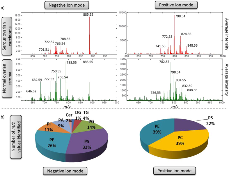
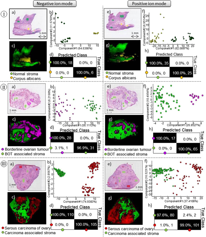
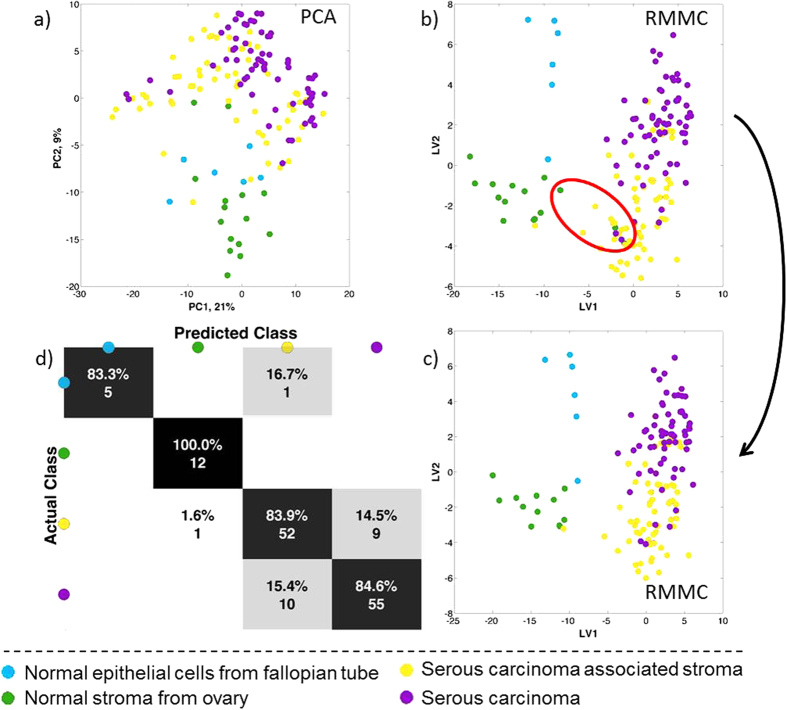
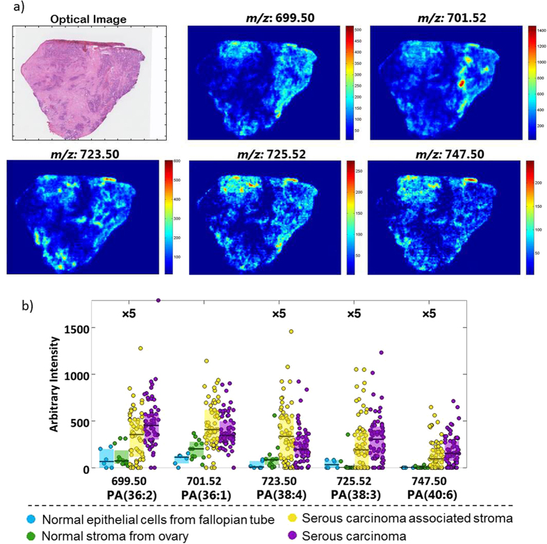
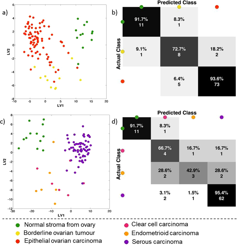
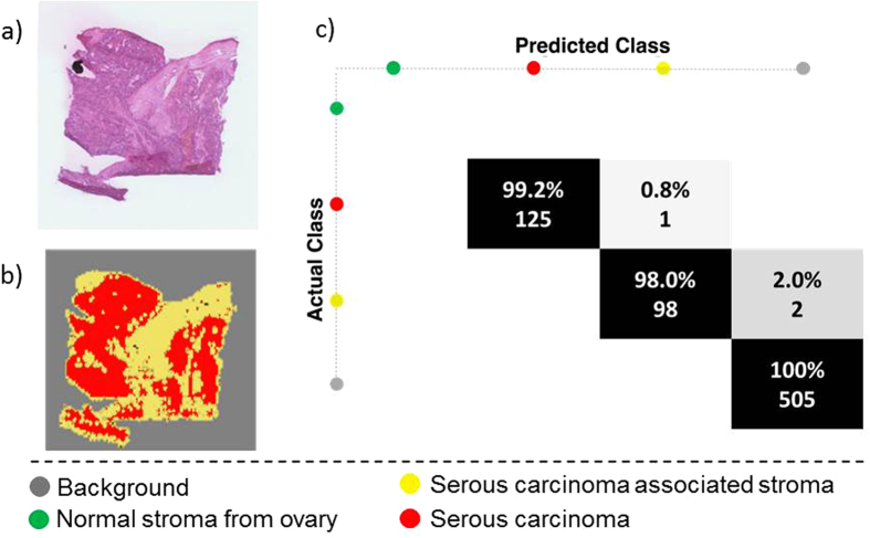
Ovarian cancer is highly prevalent among European women, and is the leading cause of gynaecological cancer death. Current histopathological diagnoses of tumour severity are based on interpretation of, for example, immunohistochemical staining. Desorption electrospray mass spectrometry imaging (DESI-MSI) generates spatially resolved metabolic profiles of tissues and supports an objective investigation of tumour biology. In this study, various ovarian tissue types were analysed by DESI-MSI and co-registered with their corresponding haematoxylin and eosin (H&E) stained images. The mass spectral data reveal tissue type-dependent lipid profiles which are consistent across the n = 110 samples (n = 107 patients) used in this study. Multivariate statistical methods were used to classify samples and identify molecular features discriminating between tissue types. Three main groups of samples (epithelial ovarian carcinoma, borderline ovarian tumours, normal ovarian stroma) were compared as were the carcinoma histotypes (serous, endometrioid, clear cell). Classification rates >84% were achieved for all analyses, and variables differing statistically between groups were determined and putatively identified. The changes noted in various lipid types help to provide a context in terms of tumour biochemistry. The classification of unseen samples demonstrates the capability of DESI-MSI to characterise ovarian samples and to overcome existing limitations in classical histopathology.


Figures

Figure 1
Figure 1
(a) Average spectra of two different samples, serous carcinoma from ovary and normal stroma from apparently normal ovary analysed in positive ion mode and negative ion mode. (b) Percentage of lipid species classes identified based on online databases.
Figure 2
Figure 2
(I) Normal ovary with two different tissue types, normal stroma (green) and corpus albicans (yellow). (II) Serous borderline tumour and (III) Serous carcinoma with two tissue types in each sample, stroma (green) and tumour tissue (red and pink). Data acquired in negative ion mode and positive ion mode. (a) and (e) Optical images and (b) and (f) PC analyses of selected regions of interest. (c) and (g) predicted RMMC components with (d) and (h) leave-one-out cross validation results.
Figure 3
Figure 3. Statistical analysis of DESI-MSI spectrum between 600–1000 Da from serous carcinoma samples with two tissue types (stroma and carcinoma) together with normal stroma from normal ovary and normal epithelium from fallopian tube.
(a) Principal component analysis (PCA) and (b) Maximum margin criteria analysis (RMMC) and (c) RMMC analysis after excluding the 3 normal appearance stroma from cancer patients with respective (d) leave one patient out cross validation using Mahalanobis as a classifier.
Figure 4
Figure 4. Characterization of phosphatidic acid class (PA) in ovarian cancer.
(a) Ion images of 5 different PA species in a serous ovarian carcinoma with (b) Box plots of the same lipid species.
Figure 5
Figure 5
Statistical analysis of DESI MS spectra between 600–1000 Da from two different perspectives: all carcinomas together, borderline ovarian tumours and normal stroma from normal ovary (a and b) and from the different carcinomas as serous, endometrioid and clear cell carcinomas together with normal stroma from normal ovary (c and d). (a and c) shows maximum margin criteria analysis (RMMC) cross validated and (b) and (d) shows leave-one-patient-out cross validation results.
Figure 6
Figure 6. Prediction of an independent sample using as training set the previous model (serous ovarian carcinoma).
(a) Optical image of the independent sample, (b) predicted image and (c) confusion matrix for classification of histopathologist-annotated tissue regions of interest.

References

    1. Menon U., Griffin M. & Gentry-Maharaj A. Ovarian cancer screening–current status, future directions. Gynecologic oncology 132, 490–495, doi: 10.1016/j.ygyno.2013.11.030 (2014). - DOI - PMC - PubMed
    1. Bast R. C. Jr., Hennessy B. & Mills G. B. The biology of ovarian cancer: new opportunities for translation. Nat Rev Cancer 9, 415–428, doi: 10.1038/nrc2644 (2009). - DOI - PMC - PubMed
    1. Roy J. E. & Hunt J. L. Detection and classification of diagnostic discrepancies (errors) in surgical pathology. Adv Anat Pathol 17, 359–365, doi: 10.1097/PAP.0b013e3181ece0db (2010). - DOI - PubMed
    1. Kommoss S. et al.. Specialized pathology review in patients with ovarian cancer: results from a prospective study. International journal of gynecological cancer: official journal of the International Gynecological Cancer Society 23, 1376–1382, doi: 10.1097/IGC.0b013e3182a01813 (2013). - DOI - PubMed
    1. Leung F., Musrap N., Diamandis E. P. & Kulasingam V. Advances in mass spectrometry-based technologies to direct personalized medicine in ovarian cancer. Advances in Integrative Medicine 1, 74–86, 10.1016/j.trprot.2013.08.001 (2013). - DOI

Publication types

MeSH terms