. 2016 Dec 15:7:13697.
doi: 10.1038/ncomms13697.

Ventral tegmental area glutamate neurons co-release GABA and promote positive reinforcement

Affiliations

Ventral tegmental area glutamate neurons co-release GABA and promote positive reinforcement

Ji Hoon Yoo et al. Nat Commun. .

Abstract

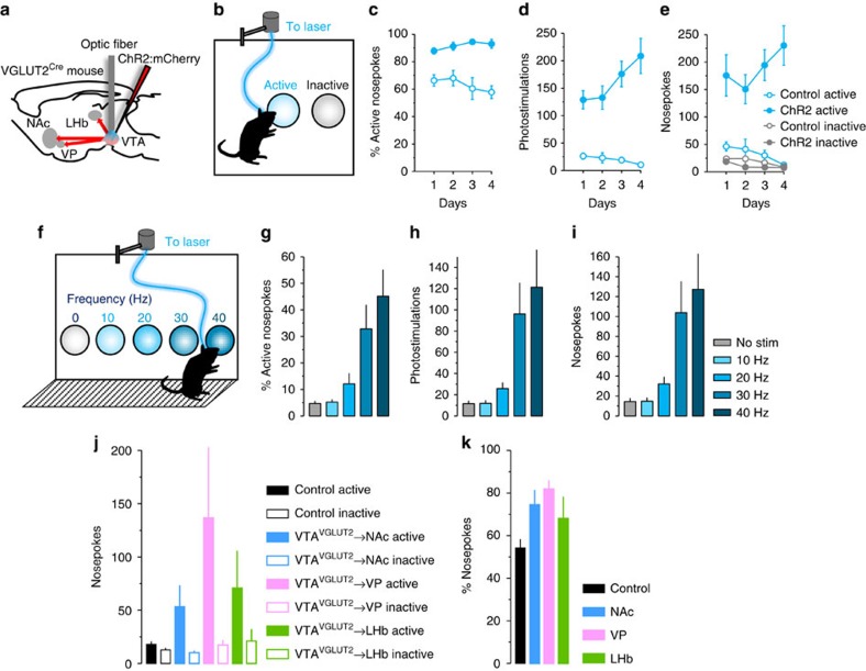
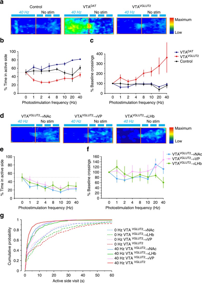
In addition to dopamine neurons, the ventral tegmental area (VTA) contains GABA-, glutamate- and co-releasing neurons, and recent reports suggest a complex role for the glutamate neurons in behavioural reinforcement. We report that optogenetic stimulation of VTA glutamate neurons or terminals serves as a positive reinforcer on operant behavioural assays. Mice display marked preference for brief over sustained VTA glutamate neuron stimulation resulting in behavioural responses that are notably distinct from dopamine neuron stimulation and resistant to dopamine receptor antagonists. Whole-cell recordings reveal EPSCs following stimulation of VTA glutamate terminals in the nucleus accumbens or local VTA collaterals; but reveal both excitatory and monosynaptic inhibitory currents in the ventral pallidum and lateral habenula, though the net effects on postsynaptic firing in each region are consistent with the observed rewarding behavioural effects. These data indicate that VTA glutamate neurons co-release GABA in a projection-target-dependent manner and that their transient activation drives positive reinforcement.


Figures

Figure 1
Figure 1. Functional expression of ChR2 in VGLUT2+ VTA neurons.
(a) Schematic illustrating strategy for selective expression of ChR2:mCherry in VGLUT2+ VTA neurons. (b) ChR2:mCherry expression (red) restricted to medial VTA in sections stained for TH (green); scales, 500 μm (top), 50 μm (bottom). (c) VTA soma clearly labelled for ChR2:mCherry were counted in each of three coronal planes and assessed for co-labeling with TH. Inset shows fraction of all ChR2:mCherry-labelled cells that co-label for TH. (d) Quantification and example images of c-Fos+ cells (green) in the VTA of control and ChR2:mCherry-expressing mice following photostimulation; P<0.01 (e) Cell-attached recordings from ChR2:mCherry+ neurons in VTA demonstrate photostimulus-induced spike entrainment and spike fidelity ≥1 for 1 s at 40 Hz. Insets show example train and mean evoked action potential waveform; blue marks indicate 5-ms light pulses.
Figure 2
Figure 2. Operant discrimination shows photostimulation of VGLUT2+ VTA neurons is positively reinforcing.
(a) Schematic illustrating strategy for selective expression and photostimulation of ChR2 in VGLUT2+ VTA neurons. (b) Schematic illustrating 2-nosepoke discrimination task where responding on the active nosepoke triggers 80 pulses at 40 Hz. (c) ChR2-expressing mice but not control mice develop a preference for the active nosepoke; P<0.001. (d) ChR2-expressing mice trigger more photostimuli than control littermates; P<0.001. (e) ChR2-expressing mice make more active nosepokes than controls; P<0.001. (f) Schematic illustrating 5-choice nosepoke task where each of 5 nosepokes are coupled to 1-s photostimulus at varying frequency. Frequency-response histograms reveal ChR2-expressing mice display an (g) ascending preference for faster stimulation; P<0.001, (h) trigger more stimuli; P<0.001, and (i) have higher response rates at nosepoke holes coupled to faster photostimulation; P<0.001 compared with responding on control nosepoke hole (for no stimulation). (j) Mice expressing ChR2 in VGLUT2+ VTA neurons were implanted instead with fibres in each of the NAc, VP or LHb to target presynaptic terminals. When placed on a restricted feeding schedule and assessed using the 2-nosepoke instrumental task, mice displayed an increase in responding; P<0.01 and a (k) preference for the active nosepoke compared with controls. Data in j and k represent average responses over 5 days.
Figure 3
Figure 3. VGLUT2+ VTA terminals co-release glutamate and GABA in projection-target restricted fashion.
Single-pulse (5-ms, blue dashes) photostimulation of (a) local VTA collaterals or (b) terminals in the NAc triggered DNQX-sensitive EPSCs (Vh=−60) but no IPSCs (Vh=0). Photostimulation of VGLUT2+ VTA terminals in (c) VP led to EPSCs in all connected cells, but also gabazine-sensitive IPSCs. All connected cells in the (d) LHb displayed both EPSCs and IPSCs. Pie charts show the percentage of neurons that showed light-triggered EPSCs only (grey), both EPSCs and IPSCs (black), or no currents (white). Bar graphs show peak amplitude of E/IPSCs, with points representing individual cells pre- and post- drug application; representative traces are inset. Scales, 50 pA, 50 ms.
Figure 4
Figure 4. VGLUT2+ VTA terminals are net excitatory in VP but inhibitory in LHb.
(a) GABA/AMPA ratios calculated from voltage-clamp recordings at Vh=0/Vh=−60 suggest the effects of GABA are predominant in the LHb compared with VP. The value of the GABA/AMPA ratio for each individual recorded neuron is represented by the grey rounds. The top insets show representative traces of GABA IPSC (black) and glutamate EPSC (grey); **P<0.01, scales 50 pA, 50 ms. (b) Representative cell-attached traces in the VP (top) or LHb (bottom) showing firing before, during (blue), and after 40-Hz 5-s photostimulation. (c) Photostimulation of VGLUT2+ VTA terminals led to a consistent increase in firing in the VP, but a decrease in the LHb. The left (black) axis and plots show mean±s.e.m. firing rates 5-s before, during (blue background), and after photostimulation; the right (grey) axis and plots show firing rates (Hz) for the individual neurons. Though we occasionally observed a transient rebound effect following light off, the firing frequency in the 5-s bin after the stimulation was not significantly different from the 5-s bin prior; ***P<0.001. (d) Data from C in 200-ms bins shows that the excitatory effects on VP and inhibitory effects on LHb persist for the duration of the 5-s stimulus train.
Figure 5
Figure 5. Mice spend less time but make more entries into compartments paired with stimulation of VTA glutamate neurons.
(a) Example location heat maps of relative times spent in 20 min RTPP 20-min RTPP assay, comparing photostimulation of ChR2-negative controls, DAT+ dopamine, or VGLUT2+ glutamate neurons in the VTA. (b) Per cent of time spent on the active side reveals a frequency-dependent increase in time spent on the active side for dopamine neuron stimulation P<0.01; low frequency stimulation of VTA glutamate neurons led to a significant reduction in time spent on active side, but this effect was mitigated with higher frequency. (c) Stimulation of VGLUT2+ VTA neurons led to a frequency-dependent increase in the number of crossings between active and inactive sides; P<0.05. (d) Example heat maps comparing photostimulation of VGLUT2+ VTA terminals in the NAc, VP or LHb. (e) Terminal stimulation leads to an apparent frequency-dependent place avoidance; P<0.001, but with (f) no decrease in crossings. (g) Significant shifts in the distributions toward shorter visits is consistent with the possibility that apparent avoidance reflects preference for brief burst stimulation rather than overt avoidance/aversion; P<0.001.
Figure 6
Figure 6. Mice prefer brief stimulation of VTA glutamate but sustained stimulation of VTA dopamine neurons.
(a) Using a 60-min 5-nosepoke choice assay with 40-Hz stimulation coupled to different stimulus durations, (b) photostimulation of VGLUT2+ VTA neurons produces a preference for short over longer stimulus duration compared with DAT+ VTA neurons; P<0.01. (c) Nosepoke responses for the varying stimulus durations; note that only the final day is shown here, but all days are displayed in Supplementary Fig. 10. (d) Using a 2-nosepoke choice assay with 40-Hz stimulation coupled to 1- or 20-s photostimulation, (e) mice expressing ChR2 in VGLUT2+ VTA neurons displayed a significant preference for shorter stimulation P<0.01. (f) In comparison, stimulation of DAT+ VTA neurons leads to the development of a preference for longer stimulus durations; P<0.001.

Similar articles

Cited by

References

    1. Fields H. L., Hjelmstad G. O., Margolis E. B. & Nicola S. M. Ventral tegmental area neurons in learned appetitive behavior and positive reinforcement. Annu. Rev. Neurosci. 30, 289–316 (2007). - PubMed
    1. Tritsch N. X., Ding J. B. & Sabatini B. L. Dopaminergic neurons inhibit striatal output through non-canonical release of GABA. Nature 490, 262–266 (2012). - PMC - PubMed
    1. Hnasko T. S. et al.. Vesicular glutamate transport promotes dopamine storage and glutamate corelease in vivo. Neuron 65, 643–656 (2010). - PMC - PubMed
    1. Stuber G. D., Hnasko T. S., Britt J. P., Edwards R. H. & Bonci A. Dopaminergic terminals in the nucleus accumbens but not the dorsal striatum corelease glutamate. J. Neurosci. 30, 8229–8233 (2010). - PMC - PubMed
    1. Tecuapetla F. et al.. Glutamatergic signaling by mesolimbic dopamine neurons in the nucleus accumbens. J. Neurosci. 30, 7105–7110 (2010). - PMC - PubMed

Publication types

MeSH terms

LinkOut - more resources