Review
. 2017 Feb;17(2):116-130.
doi: 10.1038/nrc.2016.124. Epub 2016 Dec 15.

The recurrent architecture of tumour initiation, progression and drug sensitivity

Affiliations
Review

The recurrent architecture of tumour initiation, progression and drug sensitivity

Andrea Califano et al. Nat Rev Cancer. 2017 Feb.

Abstract

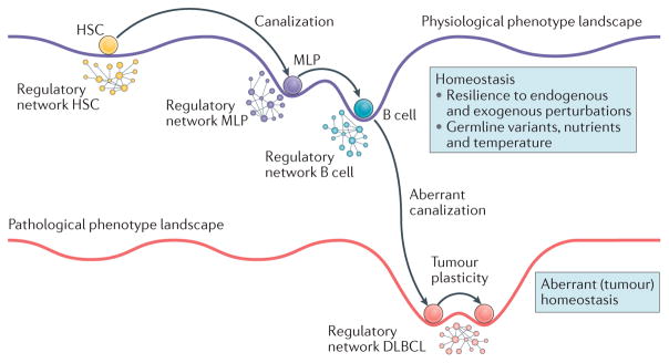
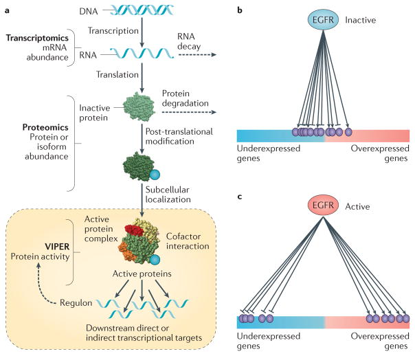
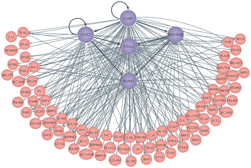
Recent studies across multiple tumour types are starting to reveal a recurrent regulatory architecture in which genomic alterations cluster upstream of functional master regulator (MR) proteins, the aberrant activity of which is both necessary and sufficient to maintain tumour cell state. These proteins form small, hyperconnected and autoregulated modules (termed tumour checkpoints) that are increasingly emerging as optimal biomarkers and therapeutic targets. Crucially, as their activity is mostly dysregulated in a post-translational manner, rather than by mutations in their corresponding genes or by differential expression, the identification of MR proteins by conventional methods is challenging. In this Opinion article, we discuss novel methods for the systematic analysis of MR proteins and of the modular regulatory architecture they implement, including their use as a valuable reductionist framework to study the genetic heterogeneity of human disease and to drive key translational applications.


Conflict of interest statement

Competing interests statement

The authors declare competing interests: see Web version for details.

Competing interests statement

A. C is founder of DarwinHealth, Inc. M. J. A. has been employed by DarwinHealth, Inc. since March 2016.

Figures

Figure 1
Figure 1. The architecture of tumour checkpoints
a | The probability densities of normal and transformed cells are shown in a principal component (PC) projection that captures most of the sample variability of four tumour types: colorectal adenocarcinoma (COAD), kidney renal clear cell carcinoma (KIRC), uterine corpus endometrial cancer (UCEC) and prostate adenocarcinoma (PRAD). These distributions show a clear single-peak structure, suggesting that the regulatory logic of the tumour cell is effective in avoiding occupancy of states that are far away from the mean. Considering that cancer tissue may also be contaminated by extensive lymphocytic and stromal cell infiltration, the variance of the normal and tumour-associated distributions is of quite comparable magnitude. A comprehensive inventory of all tumour types in The Cancer Genome Atlas (TCGA) reveals that only a handful — such as head and neck squamous cell carcinoma (HNSC), kidney renal papillary cell carcinoma (KIRP) and liver hepatocellular carcinoma (LIHC) —present with substantially greater variance than the corresponding normal tissue. b | The proposed regulatory architecture implemented by master regulator (MR) proteins in tumour checkpoints is shown. MRs (blue spheres in shaded area) represent proteins the concerted, aberrant activity of which is both necessary and sufficient for cancer cell state maintenance. Their aberrant activity is induced by genes in their upstream pathways that are mutated in a specific patient (purple spheres) selected from a larger repertoire of candidate driver genes (green spheres), the mutation of which is recurrently detected in large cohorts. Passenger mutations (pale blue spheres) that are not upstream of MRs have no effect on tumour checkpoint activity and thus on the specific phenotype that the checkpoint regulates. Arrows in this diagram show regulatory and signalling interactions, that is, how one gene product regulates other gene products. Black arrows represent crucial top-down interactions leading from patient mutations first to activation of MR proteins in the tumour checkpoint and then to activation of downstream genetic programmes that are required for tumour phenotype presentation. Grey and blue arrows represent additional regulatory interactions that do not affect and are not affected by tumour checkpoint MRs, respectively. Dashed arrows represent feedback loops implemented either between the MR layer and the upstream modulators or between genes regulated by MR proteins and upstream MR modulators. The MR protein module in the shaded area represents the tumour checkpoint. Pink spheres represent genes that are differentially expressed as a result of the aberrant activity of MR proteins in the tumour checkpoint (that is, the tumour gene expression signature). Lightning bolts represent potential therapeutic interventions using pharmacological inhibitors. Inhibiting oncoproteins mutated in a large fraction (for example, 90%)of tumour sub clones will cause relapse owing to the presence of rare, alternative subclones harbouring either alternative or bypass mutations. A bypass mutation is a mutation that activates the pathway downstream of the pharmacological intervention point. By contrast, inhibiting the tumour checkpoint may represent a more effective strategy, as it captures the effect of all upstream mutations.
Figure 2
Figure 2. Dysregulation of homeostatic control following malignant transformation and activation of dystasis control mechanisms that are responsible for the stability of tumour cell state
This figure shows how normal cell physiology is determined by the energetic landscape of its regulatory networks, enabling cells to follow specific developmental trajectories that are highly insensitive to genetic, epigenetic and environmental variability, thus achieving stable end point states. This process, also known as Waddington canalization, is illustrated in a cartoon showing differentiation from haematopoietic stem cell (HSC), to multi-lymphoid progenitor (MLP) to a fully differentiated human B cell as a set of transitions to states of progressively lower energy and thus higher stability. Disruption of this regulatory landscape by genetic alterations and environmental signals leads to physiological state loss and emergence of novel, stable disease states, for example, diffuse large B cell lymphoma (DLBCL). When multiple, quasi-isoenergetic states emerge, they can lead to coexistence of cells representing distinct tumour phenotypes in the same tumour mass or to tumour cell reprogramming to different states following treatment, a process known as tumour plasticity. For example, it has been shown that cells representing both the mesenchymal and the proneural subtype of glioma can coexist in the same tumour and that a small fraction of cells treated with tumour necrosis factor (TNF)-related apoptosis-inducing ligand (TRAIL) transiently reprogramme to a TRAIL-resistant state. Whereas normal cell homeostasis presides over the stability of physiological cell states, by making them difficult to escape, we propose that a dysregulated form of these stability control processes (that is, tumour dystasis) is responsible for the stability of tumour-associated cell states and is mechanistically implemented by a small number of master regulator (MR) proteins in a tumour checkpoint.
Figure 3
Figure 3. Diverse genetic alterations in upstream pathways contribute to aberrant NF-κB activity in DLBCL
Systematic analysis of genes in pathways upstream of the nuclear factor-κB (NF-κB) complex revealed a large repertoire of diffuse large B cell lymphoma (DLBCL)-specific genetic alterations in B cell receptor (BCR) and myeloid differentiation primary response 88 (MYD88) pathways. The presence of these mutations leads to aberrant activation of the canonical p50-RELA heterodimer and associated tumour dependency. These mutations, which are more frequent in the activated B cell (ABC) subtype of DLBCL, have provided the rationale for the clinical development of several BCR pathway inhibitors, such as ibrutinib, a Bruton tyrosine kinase (BTK) inhibitor. CARD11, caspase recruitment domain family member 11; IFN, interferon; IL, interleukin; IRAK, IL-1 receptor-associated kinase; IRF4, interferon regulatory factor 4; ITAM, immune receptor tyrosine-based activation motif; JAK1, Janus kinase 1; MALT1, mucosa-associated lymphoid tissue lymphoma translocation protein 1; PKC, protein kinase C; STAT, signal transducer and activator of transcription; TIR, Toll-interleukin receptor; TRAF6, TNF receptor associated factor 6. Adapted with permission from REF. , Nature Publishing Group.
Figure 4
Figure 4. Protein activity inference from the expression of its regulatory targets
a | Protein activity is the ultimate result of a complex cascade of molecular processes, from transcription and translation, to post-translational modification, complex formation and localization to appropriate subcellular compartments. As a result, there are no individual assays that can accurately measure protein activity in proteome-wide fashion. Instead, we have proposed that an accurate estimator of protein activity is represented by the gene expression of its transcriptional targets, that is, its regulon. This rationale is implemented by the Virtual Inference of Protein Activity by Enriched Regulon Analysis (VIPER) algorithm, based on transcription altargets inferred by reverse engineering algorithms such as Algorithm for the Accurate Reconstruction of Cellular Networks (ARACNe). b | When a protein is inactive its targets are randomly distributed in terms of differential expression. c | By contrast, when the same protein is aberrantly activated, its positively regulated targets become significantly overexpressed and its repressed targets become underexpressed. This can be effectively and quantitatively assessed by gene expression enrichment analysis methods. EGFR, epidermal growth factor receptor.
Figure 5
Figure 5. Tumour checkpoint architecture of the mesenchymal subtype of glioblastoma
Transcription factors involved in the activation of mesenchymal glioblastoma (MES-GBM) subtype are shown in purple. Overall, the six transcription factors shown in this figure—CCAAT/enhancer-binding protein-β (CEBPβ) and CEBPδ are represented by CEBP, for simplicity, as they form homodimers and heterodimers — control 74% of the genes in the mesenchymal signature of high-grade glioma. A region between 2 kb upstream and 2 kb downstream of the transcription start site of the target genes identified by Algorithm for the Accurate Reconstruction of Cellular Networks (ARACNe) was analysed for the presence of putative binding sites. When combined with analysis of gene expression profiles following short hairpin RNA (shRNA)-mediated silencing of these transcription factors, the latter were shown to bind and regulate the large majority of MES-GBM signature genes (shown in pink). In addition, CEBP (both β and δ subunits) and signal transducer and activator of transcription 3 (STAT3) were shown to regulate the other three transcription factors in the tumour checkpoint and to synergistically regulate the state of MES-GBM cells. ACTA2, actin α2; ACTN1, actinin α1; ANGPT2, angiopoietin 2; ANPEP, alanyl aminopeptidase; BACE2, β-site APP-cleaving enzyme 2; B4GALT1, β-1, 4-galactosyltransferase 1; BHLHE40, class E basic helix–loop–helix protein 40; CA12, carbonic anhydrase 12; C1QTNF1, C1q and tumour necrosis factor related protein 1; C1R, complement C1r; C1RL, complement C1r subcomponent like; CHI3L1, chitinase 3 like 1; COL4A2, collagen type IVα1 chain; ECE1, endothelin converting enzyme 1; EFEMP2, EGF containing fibulin like extracellular matrix protein 2; EFNB2, ephrin B2; EHD2, EH domain containing 2; EMP1, epithelial membrane protein 1; ESM1, endothelial cell specific molecule 1; FCGR2A, Fc fragment of IgG receptor IIa; FLNA, filamin A; FOSL2, Fos-related antigen 2; FPRL1, formyl peptide receptor-like 1; HRH1, histamine receptor H1; ICAM1, intercellular adhesion molecule 1; IFITM, interferon induced transmembrane protein; IL32, interleukin-32; TGA7, integrin subunit α7; LIF, leukaemia inhibitory factor; MMP, matrix metalloproteinase; MVP, major vault protein; MYH9, myosin heavy chain 9; MYL9, myosin light chain 9; NRP2, neuropilin 2; OSMR, oncostatin M receptor; PAPPA, pappalysin 1; PDLIM4; PDZ and LIM domain 4; PDPN, podoplanin; PELO, pelota homologue; PI3, peptidase inhibitor 3; PLA2G5, phospholipase A2 group V; PLAU, plasminogen activator, urokinase; PLAUR, PLAU receptor; PVRL2, poliovirus receptor-related 2; PTRF, polymerase I and transcript release factor; RRBP1, ribosome binding protein 1; RUNX1, runt-related transcription factor 1; SGSH, N-sulfoglucosamine sulfohydrolase; S100A11, S100 calcium binding protein A11; SLC39A8, solute carrier family 39 member 8; SOCS3, suppressor of cytokine signalling 3; TAGLN, transgelin; THBD, thrombomodulin; TIMP1, tissue inihibitor of metalloproteinase 1; TMEPAI, transmembrane prostate androgen-induced protein; TNC, tenascin C; TPP1, tripeptidyl peptidase 1; ZYX, zyxin. Adapted with permission from REF. , Nature Publishing Group.

Similar articles

Cited by

References

    1. Hoadley KA, et al. Multiplatform analysis of 12 cancer types reveals molecular classification within and across tissues of origin. Cell. 2014;158:929–944. - PMC - PubMed
    1. Shah SP, et al. The clonal and mutational evolution spectrum of primary triple-negative breast cancers. Nature. 2012;486:395–399. - PMC - PubMed
    1. Verhaak RG, et al. Integrated genomic analysis identifies clinically relevant subtypes of glioblastoma characterized by abnormalities in PDGFRA, IDH1, EGFR, and NF1. Cancer Cell. 2010;17:98–110. - PMC - PubMed
    1. Curtis C, et al. The genomic and transcriptomic architecture of 2,000 breast tumours reveals novel subgroups. Nature. 2012;486:346–352. - PMC - PubMed
    1. Cancer Genome Atlas Network. Comprehensive molecular portraits of human breast tumours. Nature. 2012;490:61–70. - PMC - PubMed

Substances