. 2016 Dec 15;11(12):e0168255.
doi: 10.1371/journal.pone.0168255. eCollection 2016.

GSK-3β/NFAT Signaling Is Involved in Testosterone-Induced Cardiac Myocyte Hypertrophy

Affiliations

GSK-3β/NFAT Signaling Is Involved in Testosterone-Induced Cardiac Myocyte Hypertrophy

Javier Duran et al. PLoS One. .

Abstract

Testosterone induces cardiac hypertrophy through a mechanism that involves a concerted crosstalk between cytosolic and nuclear signaling pathways. Nuclear factor of activated T-cells (NFAT) is associated with the promotion of cardiac hypertrophy, glycogen synthase kinase-3β (GSK-3β) is considered to function as a negative regulator, mainly by modulating NFAT activity. However, the role played by calcineurin-NFAT and GSK-3β signaling in testosterone-induced cardiac hypertrophy has remained unknown. Here, we determined that testosterone stimulates cardiac myocyte hypertrophy through NFAT activation and GSK-3β inhibition. Testosterone increased the activity of NFAT-luciferase (NFAT-Luc) in a time- and dose-dependent manner, with the activity peaking after 24 h of stimulation with 100 nM testosterone. NFAT-Luc activity induced by testosterone was blocked by the calcineurin inhibitors FK506 and cyclosporine A and by 11R-VIVIT, a specific peptide inhibitor of NFAT. Conversely, testosterone inhibited GSK-3β activity as determined by increased GSK-3β phosphorylation at Ser9 and β-catenin protein accumulation, and also by reduction in β-catenin phosphorylation at residues Ser33, Ser37, and Thr41. GSK-3β inhibition with 1-azakenpaullone or a GSK-3β-targeting siRNA increased NFAT-Luc activity, whereas overexpression of a constitutively active GSK-3β mutant (GSK-3βS9A) inhibited NFAT-Luc activation mediated by testosterone. Testosterone-induced cardiac myocyte hypertrophy was established by increased cardiac myocyte size and [3H]-leucine incorporation (as a measurement of cellular protein synthesis). Calcineurin-NFAT inhibition abolished and GSK-3β inhibition promoted the hypertrophy stimulated by testosterone. GSK-3β activation by GSK-3βS9A blocked the increase of hypertrophic markers induced by testosterone. Moreover, inhibition of intracellular androgen receptor prevented testosterone-induced NFAT-Luc activation. Collectively, these results suggest that cardiac myocyte hypertrophy induced by testosterone involves a cooperative mechanism that links androgen signaling with the recruitment of NFAT through calcineurin activation and GSK-3β inhibition.


Conflict of interest statement

The authors have declared that no competing interests exist. I confirm that my commercial affiliation does not alter our adherence to all PLOS policies on sharing data and materials.

Figures
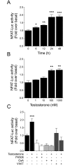
Fig 1
Fig 1. Testosterone activates NFAT in cardiac myocytes.
NFAT activity was determined in cardiac myocytes transfected with a NFAT luciferase-reporter plasmid (NFAT-Luc) and normalized relative to Renilla luciferase activity in each sample. NFAT-Luc activity is expressed as fold induction relative to non-stimulated cells. (A) Cardiac myocytes were stimulated with 100 nM testosterone for 6, 12, 24, and 48 h. (B) Concentration-dependent effect of testosterone on NFAT-Luc activity. Cells were stimulated with 1, 10, 100, or 1000 nM testosterone for 24 h. Treatment with 100 and 1000 nM testosterone significantly increased NFAT-Luc activity as compared with non-stimulated cells. (C) Pretreatment of cardiac myocytes for 30 min with FK506 (1 μM), CsA (1 μM), or 11R-VIVIT (1 μM) prior testosterone stimulation (100 nM by 24 h) reduced NFAT-Luc activation. Values are presented as the mean ± SEM (n = 5 for each condition). *p < 0.05, **p < 0.01, and ***p < 0.001 vs. control non-stimulated condition.
Fig 2
Fig 2. Testosterone-induced NFAT activation depends on androgen receptor.
Cardiac myocytes expressing NFAT-Luc were pretreated with AR inhibitors. (A) Cells were pretreated for 30 min with bicalutamide (1 mM) or cyproterone (1 μM) before stimulation with testosterone (100 nM) for 24 h. (B) Cardiac myocytes were transfected with siRNA-AR (20 nM) or non-targeting siRNA as a control. AR downregulation abolished the increase in NFAT-Luc activity induced by testosterone. Values are presented as the mean ± SEM (n = 4 for each condition). *p < 0.05 and **p < 0.01 vs. control.
Fig 3
Fig 3. Testosterone inhibits GSK-3β in cardiac myocytes.
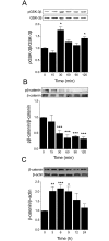
Cardiac myocytes were stimulated with 100 nM testosterone for 15, 30, 60, 90, and 120 min and subjected to Western blot analysis to determine (A) GSK-3β phosphorylation (p-GSK-3β, Ser9) and protein levels (n = 5) and (B) β-catenin phosphorylation and total β-catenin protein levels (n = 4). The densitometric analyses show the ratio of phosphorylated versus total protein. Testosterone increased GSK-3β phosphorylation at Ser9 and decreased β-catenin phosphorylation at Ser33, Ser37, and Thr41 after 30 min of stimulation. (C) Cardiac myocytes were stimulated with 100 nM testosterone for 3, 6, 9, 12, and 24 h and total β-catenin protein accumulation was determined through Western blotting (n = 5). Values are presented as the mean ± SEM. *p < 0.05, **p < 0.01, and ***p < 0.001 vs. control non-stimulated condition.
Fig 4
Fig 4. PI3K/Akt signaling is involved in GSK-3β inhibition.
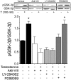
Cardiac myocytes were pretreated for 30 min with Akt-inhibitor VIII (Akti-VIII, 10 μM), LY-294002 (10 μM), or PD98059 (50 μM) prior to testosterone stimulation (100 nM) by 30 min. The densitometry results show the ratio of phosphorylated protein to total protein (n = 4 for each condition). Values are presented as the mean ± SEM. *p < 0.05 vs. control.
Fig 5
Fig 5. GSK-3β inhibition activates NFAT.
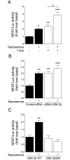
Cardiac myocytes were co-transfected with NFAT-Luc and Renilla luciferase plasmids. (A) Cells were pretreated with 1-Azk for 30 min (1 μM) prior to 100 nM testosterone stimulation for 24 h. (B) Cardiac myocytes were transfected with either siRNA-GSK-3β or a non-targeted siRNA control and then stimulated with 100 nM testosterone for 24 h. (C) Cardiac myocytes were transfected with either GSK-3βWT or GSK-3βS9A, and then stimulated with 100 nM testosterone for 24 h (n = 6 for each condition). Values are presented as the mean ± SEM. *p < 0.05, **p < 0.01, and ***p < 0.001 vs. control; # p < 0.05 vs. 1-Azk.
Fig 6
Fig 6. NFAT is involved in cardiac myocyte hypertrophy induced by testosterone.
Cardiac myocytes were pretreated with FK506 (1 μM) or CsA (1 μM) or 11R-VIVIT (1 μM) and stimulated with 100 nM testosterone for 48 h. (A) For experiments involving cellular area measurement, the cardiac myocytes were incubated with CellTracker Green and visualized by confocal microscopy (n > 80 cells from 4 independent cell cultures). (B) Protein synthesis was determined based on [3H]-leucine incorporation. The data correspond to the ratio (basal counts/min)·μg-1 protein for each experimental condition (n = 6 for each condition). Values are presented as the mean ± SEM. *p < 0.05, and ***p < 0.001 vs. control non-stimulated condition.
Fig 7
Fig 7. GSK-3β inhibition is involved in testosterone-induced cardiac myocyte hypertrophy.
Cardiac myocytes were pretreated with 1-Azk (1 μM) or transfected with siRNA-GSK-3β (A and B, respectively), or the cells were transfected with GSK-3βWT or GSK-3βS9A expression plasmid (C and D, respectively) and then stimulated with 100 nM testosterone for 48 h. For cell size measurement, cardiac myocytes were incubated with CellTracker Green and examined by confocal microscopy (n > 80 cells from 4 independent cell cultures). Protein synthesis was determined by [3H]-leucine incorporation. The data correspond to the ratio (basal counts/min)·μg-1 protein for each experimental condition (n = 6 for each condition). Values are presented as the mean ± SEM. *p < 0.05, **p < 0.01, and ***p < 0.001 vs. control non-stimulated condition.

Similar articles

Cited by

References

    1. Frey N, Katus HA, Olson EN, Hill JA. Hypertrophy of the heart: a new therapeutic target? Circulation. 2004;109(13): 1580–1589. 10.1161/01.CIR.0000120390.68287.BB - DOI - PubMed
    1. Chien KR, Knowlton KU, Zhu H, Chien S. Regulation of cardiac gene expression during myocardial growth and hypertrophy: molecular studies of an adaptive physiologic response. Faseb J. 1991;5(15): 3037–3046. - PubMed
    1. Heineke J, Molkentin JD. Regulation of cardiac hypertrophy by intracellular signalling pathways. Nat Rev Mol Cell Biol. 2006;7(8): 589–600. 10.1038/nrm1983 - DOI - PubMed
    1. Campbell SE, Farb A, Weber KT. Pathologic remodeling of the myocardium in a weightlifter taking anabolic steroids. Blood pressure. 1993;2(3):213–6. - PubMed
    1. Marsh JD, Lehmann MH, Ritchie RH, Gwathmey JK, Green GE, Schiebinger RJ. Androgen receptors mediate hypertrophy in cardiac myocytes. Circulation. 1998;98(3): 256–261. - PubMed

MeSH terms

Substances