Review
. 2017 Apr;105(4):1195-1207.
doi: 10.1002/jbm.a.35978. Epub 2017 Feb 1.

Mesenchymal stem cells in the aseptic loosening of total joint replacements

Affiliations
Review

Mesenchymal stem cells in the aseptic loosening of total joint replacements

Jukka Pajarinen et al. J Biomed Mater Res A. 2017 Apr.

Abstract

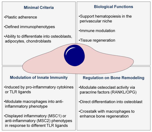
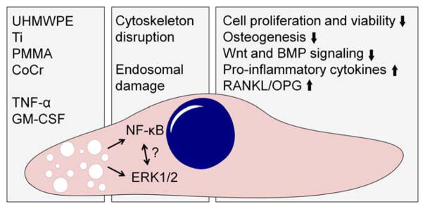
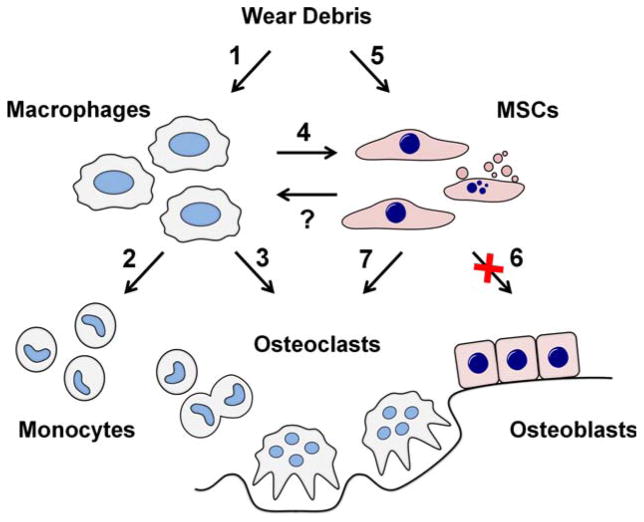
Peri-prosthetic osteolysis remains as the main long-term complication of total joint replacement surgery. Research over four decades has established implant wear as the main culprit for chronic inflammation in the peri-implant tissues and macrophages as the key cells mediating the host reaction to implant-derived wear particles. Wear debris activated macrophages secrete inflammatory mediators that stimulate bone resorbing osteoclasts; thus bone loss in the peri-implant tissues is increased. However, the balance of bone turnover is not only dictated by osteoclast-mediated bone resorption but also by the formation of new bone by osteoblasts; under physiological conditions these two processes are tightly coupled. Increasing interest has been placed on the effects of wear debris on the cells of the bone-forming lineage. These cells are derived primarily from multipotent mesenchymal stem cells (MSCs) residing in bone marrow and the walls of the microvasculature. Accumulating evidence indicates that wear debris significantly impairs MSC-to-osteoblast differentiation and subsequent bone formation. In this review, we summarize the current understanding of the effects of biomaterial implant wear debris on MSCs. Emerging treatment options to improve initial implant integration and treat developing osteolytic lesions by utilizing or targeting MSCs are also discussed. © 2017 Wiley Periodicals, Inc. J Biomed Mater Res Part A: 105A: 1195-1207, 2017.

Keywords: aseptic loosening; macrophages; mesenchymal stem cells; peri-prosthetic osteolysis; total joint replacement.


Figures

FIGURE 1
FIGURE 1
Summary of the key characteristics of MSCs. MSCs ability to modulate the innate immune response, regulate bone turnover, and directly regenerate bone make them ideal candidates for the development of cell-based therapies for aseptic loosening and other conditions were bone loss is due to chronic inflammation.
FIGURE 2
FIGURE 2
The impact of wear debris on MSCs. Biomaterial wear particles released from the implant are phagocytosed by MSCs. Particle phagocytosis induces disruption of the actin cytoskeleton and likely damage to endosomal membranes. This particle induced cell stress leads to activation of inflammatory signaling pathways, including NF-kB and ERK1/2. Activation of these pathways compromises both the viability and osteogenic differentiation ability of MSCs. Furthermore, production of selected pro-inflammatory cytokines as well as the ratio of RANKL to OPG is increased, both changes that can contribute to local osteoclast formation. In addition to direct effect of particles, also pro-inflammatory cytokines such as TNF-α and GM-CSF produced either by MSCs themselves or local macrophages likely contribute to the overall effect of wear debris. The role of Toll-like receptors and other pattern recognition receptors in the particle induced activation of inflammatory intracellular signaling pathways remain to be determined as does the detailed cross-talk between these pathways.
FIGURE 3
FIGURE 3
Role of MSCs in the aseptic loosening. Implant-derived wear debris is phagocytosed both by macrophages and MSCs. (1) Macrophages are induced to produce chemokines such as CCL2 and CCL3 as well as pro-inflammatory cytokines such as TNF-α, IL-1β and IL-6. (2) Chemokines continuously recruit additional monocytes and osteoclast precursors to the peri-implant tissue. (3) Pro-inflammatory cytokines stimulate osteoclast formation and (4) contribute to the impairment of MSC function. (5) Phagocytosed wear particles directly compromise both the viability and (6) osteogenic differentiation of MSCs. (7) Upregulation of pro-inflammatory cytokines and RANKL/OPG ratio in particle stimulated MSCs contributes to the osteoclast formation and activity. With increased osteoclast activity and suppressed osteoblast formation and function the balance of bone remodeling in the peri-prosthetic tissue turns to bone resorption with progressive peri-implant bone loss. (?) The possible effects and the therapeutic potential of MSCs reciprocally regulating macrophage function remain to be determined.

References

    1. Learmonth ID, Young C, Rorabeck C. The operation of the century: Total hip replacement. Lancet. 2007;370:1508–1519. - PubMed
    1. Pajarinen J, Lin TH, Sato T, Yao Z, Goodman S. Interaction of materials and biology in total joint replacement - successes, challenges and future directions. J Mater Chem B Mater Biol Med. 2014;2:7094–7108. - PMC - PubMed
    1. OECD. Health at a glance 2011: OECD indicators. 4.7. [Accessed on November 12th, 2016];Hip and knee replacements. 2011 :92–93. http://www.oecd.org/els/health-systems/49105858.pdf.
    1. CDC. [Accessed on November 12th, 2016];Number of all-listed procedures for discharges from short-stay hospitals, by procedure category and age. 2010 https://www.cdc.gov/nchs/data/nhds/4procedures/2010pro4_numberpro-cedure....
    1. Kurtz S, Mowat F, Ong K, Chan N, Lau E, Halpern M. Prevalence of primary and revision total hip and knee arthroplasty in the United States from 1990 through 2002. J Bone Joint Surg Am. 2005;87:1487–1497. - PubMed

LinkOut - more resources