. 2016 Dec;18(12):795-805.
doi: 10.1016/j.neo.2016.08.008.

Patient-Specific Screening Using High-Grade Glioma Explants to Determine Potential Radiosensitization by a TGF-β Small Molecule Inhibitor

Affiliations

Patient-Specific Screening Using High-Grade Glioma Explants to Determine Potential Radiosensitization by a TGF-β Small Molecule Inhibitor

N Sumru Bayin et al. Neoplasia. 2016 Dec.

Abstract

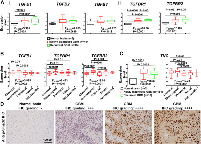
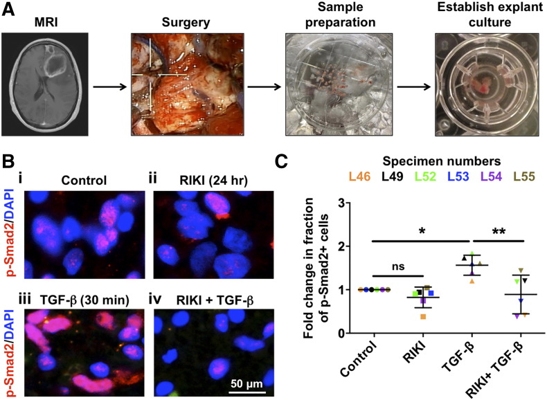
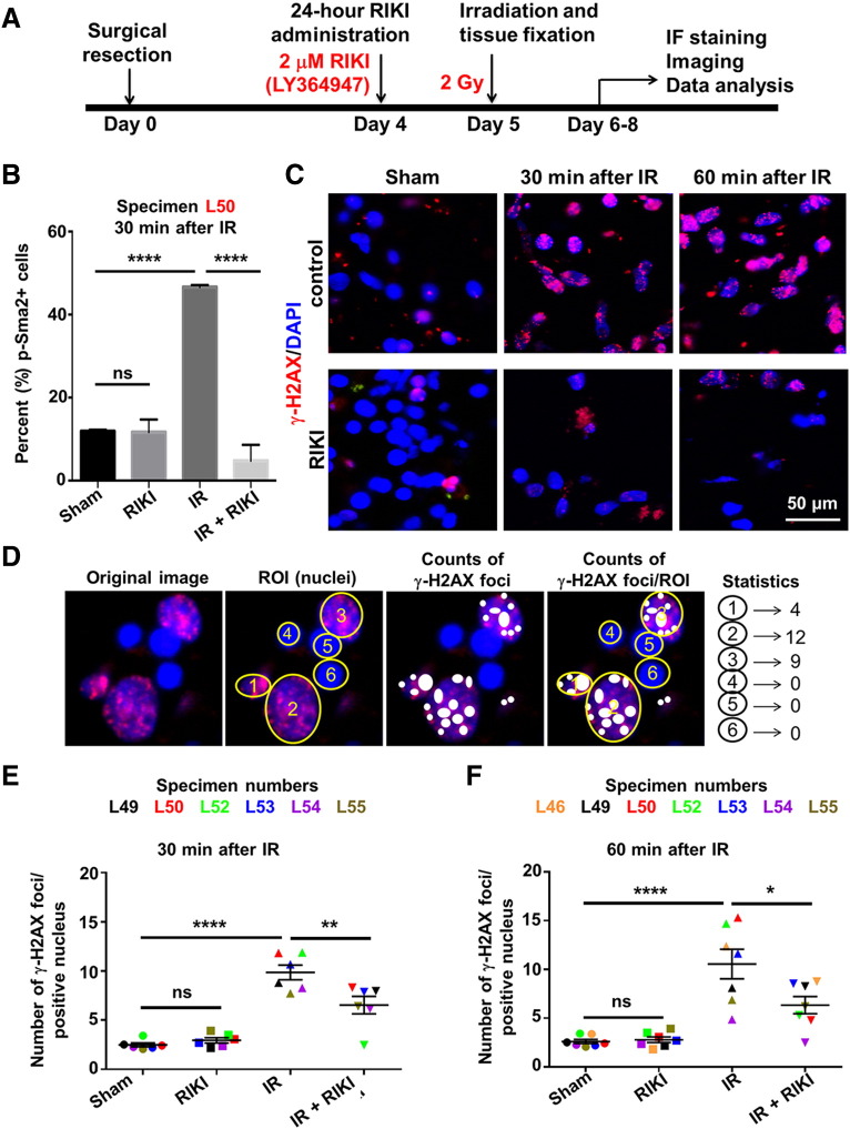
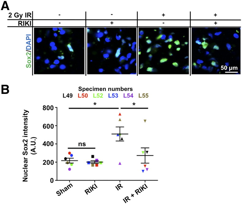
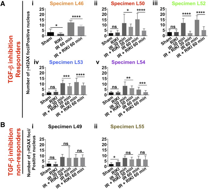
High-grade glioma (HGG), a deadly primary brain malignancy, manifests radioresistance mediated by cell-intrinsic and microenvironmental mechanisms. High levels of the cytokine transforming growth factor-β (TGF-β) in HGG promote radioresistance by enforcing an effective DNA damage response and supporting glioma stem cell self-renewal. Our analysis of HGG TCGA data and immunohistochemical staining of phosphorylated Smad2, which is the main transducer of canonical TGF-β signaling, indicated variable levels of TGF-β pathway activation across HGG tumors. These data suggest that evaluating the putative benefit of inhibiting TGF-β during radiotherapy requires personalized screening. Thus, we used explant cultures of seven HGG specimens as a rapid, patient-specific ex vivo platform to test the hypothesis that LY364947, a small molecule inhibitor of the TGF-β type I receptor, acts as a radiosensitizer in HGG. Immunofluorescence detection and image analysis of γ-H2AX foci, a marker of cellular recognition of radiation-induced DNA damage, and Sox2, a stem cell marker that increases post-radiation, indicated that LY364947 blocked these radiation responses in five of seven specimens. Collectively, our findings suggest that TGF-β signaling increases radioresistance in most, but not all, HGGs. We propose that short-term culture of HGG explants provides a flexible and rapid platform for screening context-dependent efficacy of radiosensitizing agents in patient-specific fashion. This time- and cost-effective approach could be used to personalize treatment plans in HGG patients.


Figures

Figure 1
Figure 1
Heterogeneous activation of TGF-β signaling in HGGs. (A) Analysis of TCGA data on expression levels of five transcripts related to TGF-β signaling in GBM specimens (i: TGF-β ligands, ii: TGF-β receptors). For each transcript, expression levels across three different groups, normal brain (black), newly diagnosed GBM (red), and recurrent GBM (green), have been plotted as box-whisker plots. (B) Expression levels of transcripts TGFB1, TGFBR1, and TGFBR2 across different molecular subgroups of GBM. (C) Analysis of TNC mRNA levels, a downstream target of TGF-β signaling, in normal brain (black), newly diagnosed GBM (red), and recurrent GBM (green). ANOVA statistics with post hoc comparisons were used in A to C. (D) Representative images of the grading system used to analyze p-Smad2 immunostaining in normal brain and HGG specimens.
Figure 2
Figure 2
Establishment of HGG cultures and effects of RIKI on TGF-β–induced Smad2 phosphorylation. (A) Schematic showing the establishment of cultures from surgical HGG specimens. (B) Representative images of p-Smad2 (red) immunofluorescence analysis (specimen L46) at baseline (i), after 24 hours of RIKI (ii), after 30 minutes of TGF-β (iii), and after combined RIKI and TGF-β (iv). Nuclei are counterstained with DAPI (blue). RIKI was given for 24 hours and TGF-β for 30 minutes prior to assays. (C) The fraction of p-Smad2–positive cells increased after TGF-β stimulation in GBM cultures. This effect was blocked by pretreatment with RIKI. The mean values and standard error of six specimens are shown [AVOVA F(3,20) = 8.876, P = 0.006]. *P < 0.05; **P < 0.01.
Figure 3
Figure 3
DSB recognition dynamics in HGG explant cultures analyzed by γ-H2AX immunoreactivity. (A) Experimental timeline for testing radiosensitizing effects of TGF-β inhibition. (B) RIKI prevented the radiation-induced increase in the percentage of p-Smad2+ cells in explants from specimen L50. RIKI had no effect on the fraction of p-Smad2+ cells in the absence of radiation [ANOVA F(3,8) = 170.3, P < 0.0001]. (C) Representative images of effects of RIKI on γ-H2AX (red) immunofluorescence (specimen L46) before and after radiation. Nuclei are counterstained with DAPI (blue). (D) Schematic representation of quantitation of γ-H2AX foci. Only the positive nuclei were considered for analysis. (E) Cumulative data for γ-H2AX foci/positive nucleus in six explants 30 minutes after radiation [ANOVA F(3,20) = 32.38, P < 0.0001]. (F) Collective data for γ-H2AX foci/positive nucleus in seven explants 60 minutes after irradiation [ANOVA F(3,24) = 17.44, P < 0.0001]. ns, not significant. *P < 0.05; **P < 0.01; ****P < 0.0001.
Figure 4
Figure 4
Inhibition of TGF-β signaling prevents radiation-induced upregulation of GSC marker Sox2. (A) Representative images showing effects of RIKI on Sox2 immunostaining (green) before and after radiation (specimen L52). Nuclei were counterstained with DAPI (blue). (B) Nuclear Sox2 intensity at 60 minutes after 2-Gy irradiation in six HGG explant cultures [ANOVA F(3,20) = 5.837, P = 0.0049]. *P < 0.05.
Figure 5
Figure 5
The response to TGF-β inhibition varies among different specimens. (Ai-v) Five explants responded to RIKI with decreases in radiation-induced γ-H2AX foci. (i) L46 [ANOVA F(3116) = 186.9, P < 0.0001]. (ii) L50 [ANOVA F(5174) = 41.49, P < 0.0001]. (iii) L52 [ANOVA F(5174) = 91.12, P < 0.0001]. (iv) L53 [ANOVA F(5174) = 84.59, P < 0.0001]. (v) L54 [ANOVA F(5174) = 43.16, P < 0.0001]. (Bi-ii) In two explants, RIKI did not suppress radiation-induced γ-H2AX foci. (i) L49 [ANOVA F(5174) = 12.93, P < 0.0001]. (ii) L55 [ANOVA F(5174) = 27.70, P < 0.0001]. *P < 0.05; **P < 0.01; ***P < 0.001; ****P < 0.0001.

References

    1. Stupp R, Mason WP, van den Bent MJ, Weller M, Fisher B, Taphoorn MJ, Belanger K, Brandes AA, Marosi C, Bogdahn U. Radiotherapy plus concomitant and adjuvant temozolomide for glioblastoma. N Engl J Med. 2005;352:987–996. - PubMed
    1. Gilbert MR, Dignam JJ, Armstrong TS, Wefel JS, Blumenthal DT, Vogelbaum MA, Colman H, Chakravarti A, Pugh S, Won M. A randomized trial of bevacizumab for newly diagnosed glioblastoma. N Engl J Med. 2014;370:699–708. - PMC - PubMed
    1. Verhaak RG, Hoadley KA, Purdom E, Wang V, Qi Y, Wilkerson MD, Miller CR, Ding L, Golub T, Mesirov JP. Integrated genomic analysis identifies clinically relevant subtypes of glioblastoma characterized by abnormalities in PDGFRA, IDH1, EGFR, and NF1. Cancer Cell. 2010;17:98–110. - PMC - PubMed
    1. Eckel-Passow JE, Lachance DH, Molinaro AM, Walsh KM, Decker PA, Sicotte H, Pekmezci M, Rice T, Kosel ML, Smirnov IV. Glioma groups based on 1p/19q, IDH, and TERT promoter mutations in tumors. N Engl J Med. 2015;372:2499–2508. - PMC - PubMed
    1. Singh SK, Hawkins C, Clarke ID, Squire JA, Bayani J, Hide T, Henkelman RM, Cusimano MD, Dirks PB. Identification of human brain tumour initiating cells. Nature. 2004;432:396–401. - PubMed

MeSH terms