Review
. 2017 Mar;27(3):172-188.
doi: 10.1016/j.tcb.2016.11.003. Epub 2016 Dec 13.

Extracellular Vesicles: Unique Intercellular Delivery Vehicles

Affiliations
Review

Extracellular Vesicles: Unique Intercellular Delivery Vehicles

Sybren L N Maas et al. Trends Cell Biol. 2017 Mar.

Abstract

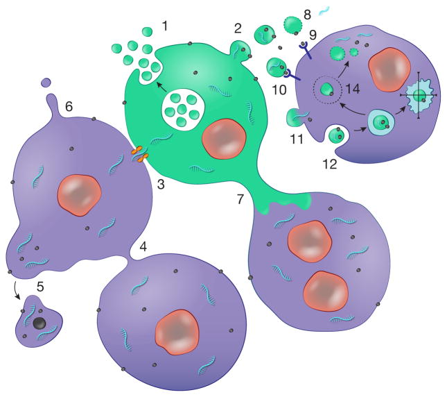
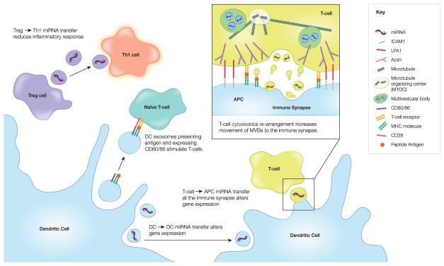
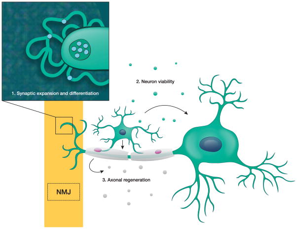
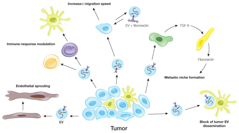
Extracellular vesicles (EVs) are a heterogeneous collection of membrane-bound carriers with complex cargoes including proteins, lipids, and nucleic acids. While the release of EVs was previously thought to be only a mechanism to discard nonfunctional cellular components, increasing evidence implicates EVs as key players in intercellular and even interorganismal communication. EVs confer stability and can direct their cargoes to specific cell types. EV cargoes also appear to act in a combinatorial manner to communicate directives to other cells. This review focuses on recent findings and knowledge gaps in the area of EV biogenesis, release, and uptake. In addition, we highlight examples whereby EV cargoes control basic cellular functions, including motility and polarization, immune responses, and development, and contribute to diseases such as cancer and neurodegeneration.

Keywords: exosomes; extracellular vesicles; microvesicles.


Figures

Fig. 1
Fig. 1. Cellular sharing of macromolecular information
Cells have a number of ways of exchanging molecules which are facilitated by being maintained within a membrane boundary. These include deployment of EVs by: 1) release of exosomes through fusion of MVBs with the plasma membrane, and 2) budding of microvesicles off the plasma membrane. 3) In addition, cells in physical contact can form gap junctions allowing exchange of small molecules, including miRNAs. Other modes include: 4) connection of cells through nanotubes; 5) blebbing off of larger vesicles, especially from cancer cells e.g. oncosomes; 6) formation of membrane protrusions which release vesicles from their tips; and 7) larger diameter microtubes connecting cells. In the case of EVs there are a number of ways for information transfer: 8) lysis of vesicles in the extracellular space releasing their contents, including 9) free ligands and 10) ligands on the surface of vesicles, which stimulate receptors on the cell surface. Uptake of EV cargo can occur through: 11) fusion of the vesicle with the plasma membrane or 12) uptake by different types of endocytosis. In the latter case the fate of the vesicle and its content can be: 13) progression through the degradative pathway to lysosomes; and/or 14) escape from the endosome compartment to release contents into the cell cytoplasm where they may be functional. References for these pathways are given in the text.
Fig. 2
Fig. 2. EV-mediated communication in the immune system
EV exchange is an important part of the communication between immune cells. In particular, T cells have been shown to both receive and deliver messages via EVs. Thus, peptide-containing MHC on EVs can stimulate naive T cells. In addition, miRNAs transferred in T-cell derived EVs can influence recipient cell gene expression. A critical structure where much of this takes place is the immune synapse, a cell-cell adhesion structure characterized by cytoskeletal reorganization. Both shed EVs containing the T cell receptor (TCR) and exosomes carrying miRNAs are transferred at the immune synapse.
Fig. 3
Fig. 3. EV-mediated communication in the nervous system
Neurons communicate with a variety of cells via EVs: 1) At the neuromuscular junction (NMJ), neuron-derived EVs promote synaptic expansion and differentiation; 2) In the central nervous system, oligodendrocyte-derived EVs promote neuronal viability and firing rate; and 3) In the peripheral nervous system, Schwann cell EVs promote axon regeneration.
Fig. 4
Fig. 4. Role of EVs in cancer
Cancer-derived EVs influence both stromal and tumor cells. These EVs can induce endothelial sprouting and neovascularization. Incubation of cancer-EVs with T-cells leads both to apoptosis in CD8+ T-cells and the expansion of CD4+ towards T-regulatory cells. Monocytes differentiate towards a more tumor supportive phenotype after incubation with cancer-EVs. As cancer cells migrate, EVs from tumor cells can accelerate this process. EVs from malignant cancer cells induce less-malignant cells to migrate faster; however, the mechanism for this is unknown. Tumor cells load fibronectin onto EVs in an autocrine manner facilitating adhesion formation and rate of migration. Tumor-derived EVs can also set up metastatic niches at a variety of locations. In the liver, cancer-EVs induce TGF-beta production by Kupffer cells, which promotes fibronectin production by hepatic stellate cells. This fibrotic environment enhances retention of neutrophils and macrophages in the liver creating a favorable metastatic niche. In some cases, local cells may counter the effect of cancer-EVs, such as subcapsular macrophages that limit dissemination of cancer-EVs from lymph nodes.

Similar articles

Cited by

References

    1. Cocucci E, et al. Shedding microvesicles: artefacts no more. Trends Cell Biol. 2009;19(2):43–51. doi: 10.1016/j.tcb.2008.11.003. - DOI - PubMed
    1. Valadi H, et al. Exosome-mediated transfer of mRNAs and microRNAs is a novel mechanism of genetic exchange between cells. Nat Cell Biol. 2007;9:654–659. - PubMed
    1. Skog J, et al. Glioblastoma microvesicles transport RNA and protein that promote tumor growth and provide diagnostic biomarkers. Nat Cell Biol. 2008;10(12):1470–1476. doi: 10.1038/ncb1800. - DOI - PMC - PubMed
    1. Tkach M, Théry C. Communication by extracellular vesicles: Where we are and where we need to go. Cell. 2016;164(6):1226–1232. doi: 10.1016/j.cell.2016.01.043. - DOI - PubMed
    1. Abels ER, Breakefield XO. Introduction to extracellular vesicles - biogenesis, RNA cargo selection, content, release, and uptake. Cell Mol Neurobiol. 2016;36(3):301–312. doi: 10.1007/s10571-016-0366-z. - DOI - PMC - PubMed

Publication types