Review
. 2017 Jan-Feb;120(1-2):78-95.
doi: 10.1016/j.ymgme.2016.11.007. Epub 2016 Nov 29.

Mucopolysaccharidosis IVA and glycosaminoglycans

Affiliations
Review

Mucopolysaccharidosis IVA and glycosaminoglycans

Shaukat Khan et al. Mol Genet Metab. 2017 Jan-Feb.

Abstract

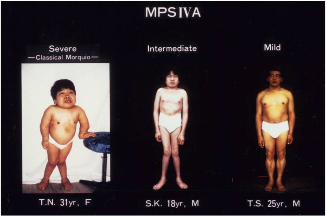
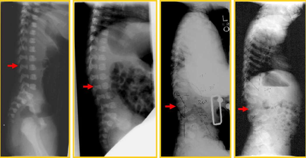
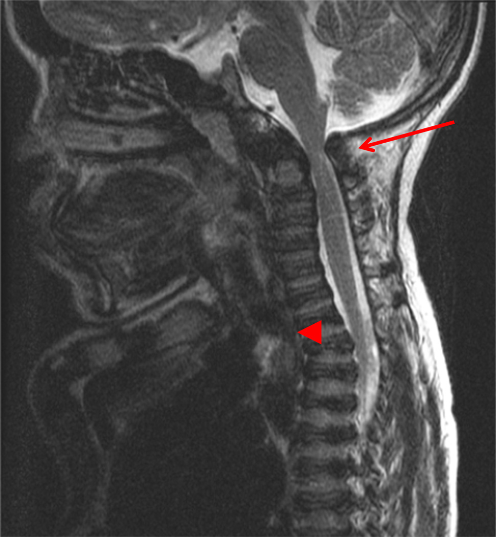
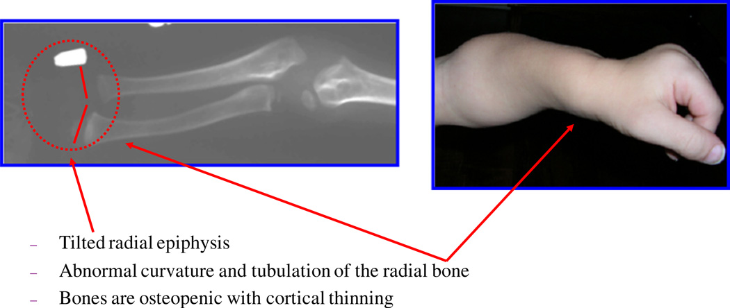
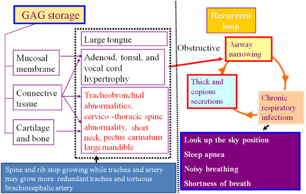
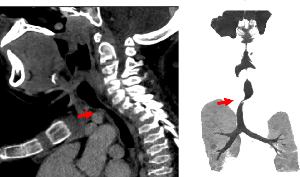
Mucopolysaccharidosis IVA (MPS IVA; Morquio A: OMIM 253000) is a lysosomal storage disease with an autosomal recessive trait caused by the deficiency of N-acetylgalactosamine-6-sulfate sulfatase. Deficiency of this enzyme leads to accumulation of specific glycosaminoglycans (GAGs): chondroitin-6-sulfate (C6S) and keratan sulfate (KS). C6S and KS are mainly produced in the cartilage. Therefore, the undegraded substrates are stored primarily in cartilage and in its extracellular matrix (ECM), leading to a direct impact on cartilage and bone development, and successive systemic skeletal dysplasia. Chondrogenesis, the earliest phase of skeletal formation, is maintained by cellular interactions with the ECM, growth and differentiation factors, signaling pathways, and transcription factors in a temporal-spatial manner. In patients with MPS IVA, the cartilage is disrupted at birth as a consequence of abnormal chondrogenesis and/or endochondral ossification. The unique skeletal features are distinguished by a disproportional short stature, odontoid hypoplasia, spinal cord compression, tracheal obstruction, pectus carinatum, kyphoscoliosis, platyspondyly, coxa valga, genu valgum, waddling gait, and laxity of joints. In spite of many descriptions of these unique clinical features, delay of diagnosis still happens. The pathogenesis and treatment of systemic skeletal dysplasia in MPS IVA remains an unmet challenge. In this review article, we comprehensively describe historical aspect, property of GAGs, diagnosis, screening, pathogenesis, and current and future therapies of MPS IVA.

Keywords: Chondroitin-6-sulfate; Keratan sulfate; Mucopolysaccharidosis IVA; N-acetylgalactosamine-6-sulfate sulfatase; Skeletal dysplasia.


Conflict of interest statement

Conflict of interestAll the authors contributed to the Review Article and had no conflict of interest with any other party.Shaukat Khan, Carlos J. Alméciga-Díaz, Kazuki Sawamoto, William G. Mackenzie, Mary C. Theroux, Christian Pizarro, Robert W. Mason, Tadao Orii, and Shunji Tomatsu declare that they have no conflict of interests.

Figures

Fig. 1
Fig. 1
Clinical pictures of MPS IVA patients (adapted from Educational CD for Morquio and permitted by Carol Ann Foundation). Left panel; this is 31 year old female patient with a severe form of MPS IVA. She is 90 cm tall, showing skeletal deformities including pectus carinatum, bilateral genu valgum, diffuse corneal clouding, atlantoaxial subluxation, and hyperlaxity of joints. The genotype is c.1288_1289delCA/c.1288_1289delCA Middle panel; this is 18 year old male patient with an intermediate form of disease. He is 135 cm tall with the milder skeletal deformities of pectus carinatum, bilateral genu valgum, and hyperlaxity of joints and corneal clouding. The genotype is p.R94G/p.N204K. Right panel; this is 25 year old male patient with a mild form of disease. He is 157 cm tall and has milder skeletal deformities of thoracolumbar gibbus, mild platyspondyly and anterior wedging of the bodies in X-rays but no genu valgum. The genotype is pN204K/pN204K. All three patients have normal intelligence.
Fig. 2
Fig. 2
Lateral view of progressive changes of spine with age in a patient (adapted from Educational CD for Morquio and permitted by Carol Ann Foundation). 1 day; a sacral dimple was noted at the delivery and the suspected anterior beaking at the level of L1. It could also be seen in normal baby. 2 months; the anterior beaking was more prominent at the level of L1 with kyphosis. 15 months; unusual kyphosis in the thoracic spine and the gibbus at L1 were prominent with the anterior beaking. L1 vertebral body was compressed. The flaring of the anterior lateral ribs was observed. 32 months; the accentuated dorsal thoracolumbar kypholordosis with the gibbus deformity was noted remarkably. The advanced platyspondylia, irregularity, and anterior bealdng of vertebral bodies are characteristic of MPS IVA.
Fig. 3
Fig. 3
MRI of cervical spine in a 13 years patient. The arrow shows C1-C2 spinal cord compression and the arrow head specifies tracheal obstruction. A baseline study of the upper cervical anatomy is recommended no later than 2 years or at diagnosis using flexion/extension X-ray films. If severe pain or pain associated with weakness of strength or tremors (or clonus) in the arms or legs occur, the patient should have studies of the neck to evaluate for the slippage (subluxation) of the neck vertebrae and compression of the spinal cord.
Fig. 4
Fig. 4
Characteristic bone deformity in the upper extremity (adapted from Educational CD for Morquio and permitted by Carol Ann Foundation). The epiphyseal involvement characteristic of MPS IVA is exemplified by the tapered irregular distal radius and ulna. The bones are osteopenic with cortical thinning. Upper extremities in a child aged 2 years 3 months (left panel). Note the irregular epiphyses and widened metaphyses. Cortical thinning and mild widening of the diaphysis of the humerus are visible. With age, the bone deformity progresses, e.g. with tilting of the radial epiphysis towards the ulna (10 years old; right panel). The humerus usually appears shortened later.
Fig. 5
Fig. 5
Hand deformity in MPS IVA (adapted from Educational CD for Morquio and permitted by Carol Ann Foundation). The radiograph in a 6-year-old patient shows the tapering of the proximal portion of metacarpals 2 through 5 and small irregular carpal bones. The joints may become hyperlaxity by the age of 2 years. The hands with age take on a characteristic with tilting of the radial epiphysis towards the ulna, resulting from a combination of metaphyseal deformities, hypoplasia of the bones, and degradation of connective tissues near the joint secondary to GAG accumulation.
Fig. 6
Fig. 6
Hip joint deformity. The radiograph of a 6-year-old patient shows bone abnormality in lower extremities. Multiple abnormalities are present in the pelvis, including spondyloepiphyseal dysplastic femoral heads and oblique acetabular roof with coxa valgus deformity and flared iliac wings.
Fig. 7
Fig. 7
ERT on Morquio A mice at 8 weeks old treated for 12 (upper) and 24 (lower) weeks with 250 U/g of E6-GALNS. Light microscopy of growth plates in femur from untreated, E6 GALNS-treated, or non-tagged (native) enzyme. An untreated mouse shows marked storage material in chondrocytes (A and D). Vacuolated chondrocytes are obvious. Chondrocytes showed a slight response after 12 and 24 weeks of treatment with native enzyme (C and F). The cartilage cells treated with bone-targeting enzymes have substantial reduction of storage material after 24 weeks of treatment (E). ERT, enzyme-replacement therapy; GALNS, N-acetylgalactosamine-6-sulfate sulfatase.
Fig. 8
Fig. 8
Pathophysiology of difficult airway in Morquio A patients. Both restrictive and obstructive respiratory pathology are common in Morquio A patients. The restrictive defect is due to thoracic cage deformity, and the obstructive defect is from tracheobronchial abnormalities, large tongue and mandible, adenoidal, tonsillar, and vocal cord hypertrophy by accumulation of storage materials. Imbalance of growth between trachea, cervical spine, and brachiocephalic artery causes tracheal obstruction. Moreover, Morquio A patients have small nasal passages caused by thickened mucous membranes and thick and copious secretions. Chronic upper respiratory tract infection further decrease the already diminished airway lumen (adapted from Educational CD for Morquio and permitted by Carol Ann Foundation).
Fig. 9
Fig. 9
Tracheal obstruction. CT in a 13-year-old patient shows a severe tracheal obstruction. Tracheal narrowing, often due to impression from the crossing bracheocephalic (innominate) artery, increases with age. Note the position of the brachiocephalic artery anterior to the trachea (arrows). Cervico-thoracic spine moves forward while a severe pectus carinatum compresses backward.

References

    1. Morquio L. Sur une forme de dystrophic osseuse familiale. Arch. Med. Infant. 1929;32:129–135.
    1. JF B. Chondro-osteo-dystrophy. Roentgenographic and clinical features of a child with dislocation of vertebrae. Am. J. Surg. 1929;7:404–410. - PubMed
    1. Pedrini V, Lennzi L, Zambotti V. Isolation and identification of keratosulphate in urine of patients affected by Morquio-Ullrich disease. Proc. Soc. Exp. Biol. Med. 1962;110:847–849. - PubMed
    1. McKusick VA, Kaplan D, Wise D, Hanley WB, Suddarth SB, Sevick ME, Maumanee AE. The genetic mucopolysaccharidoses. Medicine. 1965;44:445–483. - PubMed
    1. Orii T, Minami R, Chiba T, Yamaguchi M, Tsugawa S, Nakao T, Horino K, Sakuma K. Study on Morquio syndrome bone metabolism (in Japanese) 1971;5:72–78.

Publication types

Substances